More melanoma screening won’t close the racial gap in survival

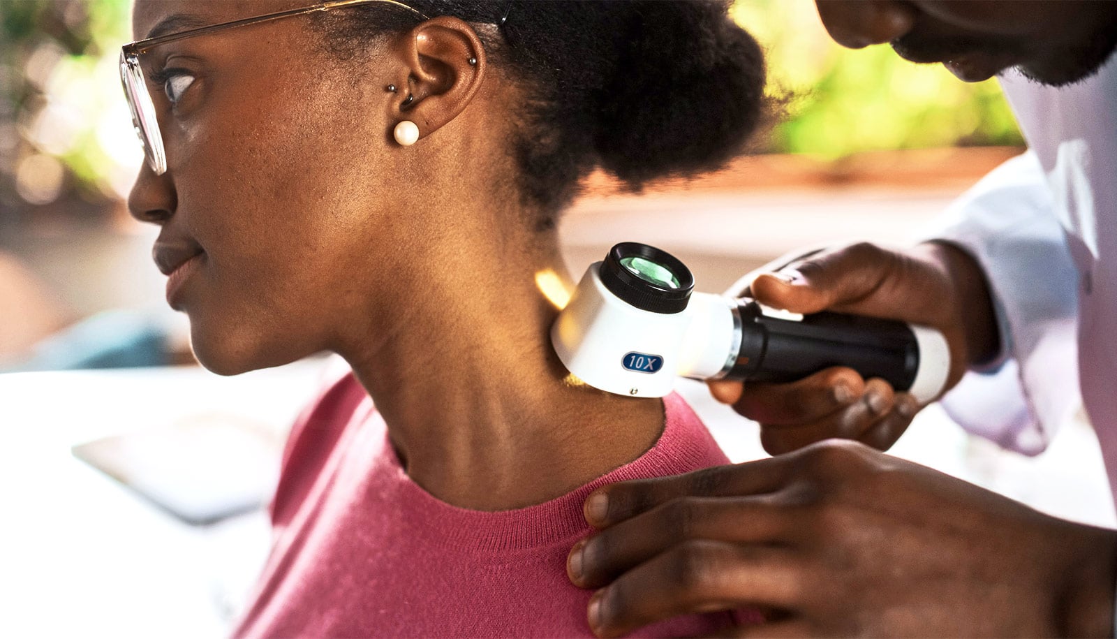
Increased skin cancer screening in people with skin of color is not sufficient to address racial disparities in melanoma survival rates, according to a new study.

Melanoma causes the most deaths of any skin cancer but is usually treatable if caught early. Although the disease is most common in white people, survival odds are worse in people with darker skin tones.

“In this study, we asked whether screening could address this disparity by helping detect melanoma early,” says senior author Laura Ferris, a dermatologist at the University of Pittsburgh Medical Center and professor of dermatology at the Pitt School of Medicine.

“Our findings suggest that regular skin checks are not the answer, but that doesn’t mean that we should be offering less care or that our work is done. We need to investigate other approaches to improve outcomes for melanoma in patients with skin of color.”

For the study, published in JAMA Dermatology, Ferris and her team analyzed data from 60,680 patients who self-reported as Hispanic, Alaska Native, American Indian, Asian, Black, or Pacific Islander. Of those, 12,738 were screened for skin cancer, and 47,942 were not screened.

During the five-year study period, only eight melanomas were detected in this population, and just one of these was identified during a screening visit. Four were identified by health care professionals during other types of visits and three were detected by the patient or a family member.

The results suggest that to detect one melanoma case in racial and ethnic minority populations, more than 12,000 screenings need to be done. For comparison, the number needed to screen in white patients is 373, the researchers found in an earlier study.

“This is an almost unfathomable number of doctor’s visits to find one melanoma,” says Ferris. “Rather than screening everyone, educating physicians about presentation of melanoma in skin of color, educating the public about their risk of melanoma, and making sure that people have access to a dermatologist when they have a suspicious lesion could be more effective in improving early detection.”

While people often think of melanomas as being sun-induced, not all forms of the disease are caused by sun exposure. Certain types of melanomas can arise on the palms of the hand, soles of the feet, and places that are always covered by clothes, and these tend to be more common in people with darker skin.

“UV exposure is the biggest modifiable risk factor for melanoma, so sun protection is incredibly important, but it’s not the only factor,” says Ferris. “If you have a suspicious lesion somewhere that is always covered by a shirt, it could still be melanoma. We encourage patients to seek care regardless of their perceived risk.”

Beyond early detection, better treatments for melanoma could also help address disparities in survival rates, Ferris says. Most of the current therapies for the disease were tested in non-diverse, mostly white populations, so it’s important that future clinical trials include diverse participants.

Additional coauthors are from Drexel University and the University of Pittsburgh.

The University of Pittsburgh Melanoma and Skin Cancer Program funded the work.

Source: University of Pittsburgh

source

Expert: No need to panic about leprosy in Florida

Cases of leprosy are increasing in Florida. Dermatologist Andrea Maderal puts that information into reassuring context.

The United States Centers for Disease Control and Prevention (CDC) has issued a new report this week warning that the central part of the state is seeing the majority of cases statewide, and one-fifth of all new cases across the country.

And unlike previous cases of leprosy—also known as Hansen’s disease—researchers are still trying to determine how exactly patients are getting the infection.

While in the past, patients diagnosed with leprosy had often traveled to another country where the disease is more prevalent, or they had some contact with the nine-banded armadillo—known to carry the bacteria that causes leprosy—a recent case has not been tied to those two factors. This prompted CDC officials to conclude that the bacteria causing leprosy may have become endemic, or it may now be found naturally in Florida.

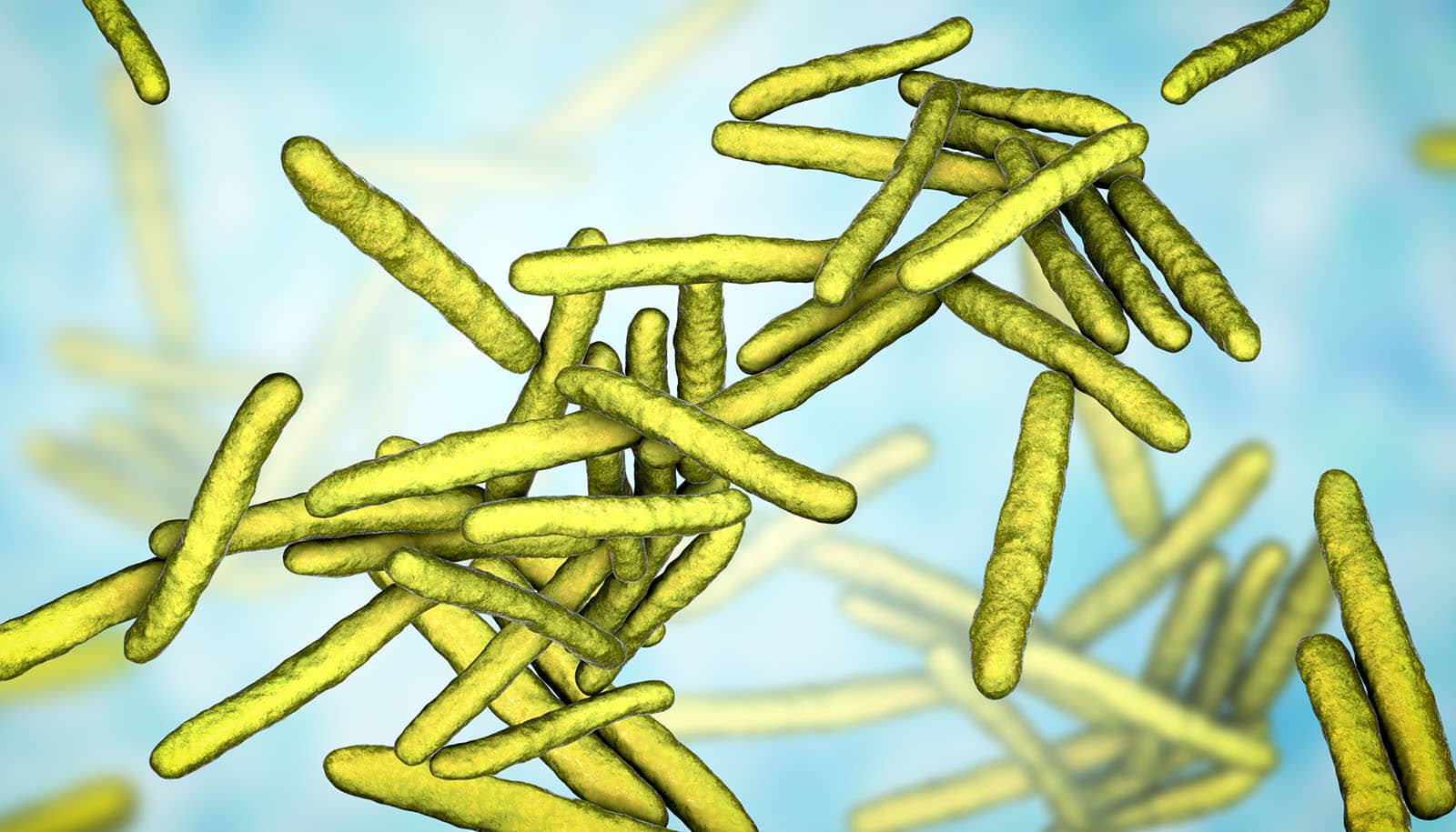
Hansen’s disease is an infection caused by a bacteria from the Mycobacterium leprae complex that primarily affects the skin and peripheral nervous system, the CDC reports. Those who contract the infection often have a skin rash that typically involves a loss of sensation, says Maderal, an assistant professor of dermatology at the University of Miami Miller School of Medicine and director of the University of Miami/Jackson Memorial Hospital Hansen’s Disease Program, which is one of 16 federally funded programs across the nation. To confirm that the rash is leprosy, typically dermatologists need to do a skin biopsy, Maderal points out.

And while headlines on Tuesday suggested a dangerous surge in cases, Maderal cautions that Hansen’s disease is still extremely rare, and it is treatable. Annually, only about 150 people get Hansen’s disease in the US and about 200,000 people are diagnosed across the globe.

Here, Maderal addresses questions about the outbreak and the disease:

source

Brain imaging could lead to earlier schizophrenia diagnosis

New research has identified age-related changes in brain patterns associated with the risk for developing schizophrenia.

The discovery could help clinicians identify the risk for developing mental illness earlier and improve treatment options.

The study appears in the Proceedings of the National Academy of Sciences.

The researchers used a hybrid, data-driven method called Neuromark to extract reliable brain networks from the neuroimaging data which were then further analyzed in the study.

The researchers started with functional MRI scans (fMRI) to detect age-related changes in brain connectivity and their association with schizophrenia risk. The research identified high-risk individuals for developing psychosis during late adolescence and early adulthood.

Using this novel approach to existing functional neuroimaging datasets led to a breakthrough in understanding both genetic and clinical risks for schizophrenia in the context of how brain regions communicate with each other.

“This study combined over 9,000 data sets using an approach which computes functional brain networks adaptively while also allowing us to summarize and compare across individuals,” says Vince Calhoun, director of the Tri-institutional Center for Translational Research in Neuroimaging and Data Science (TReNDS) center based at Georgia State University.

“This led us to a really interesting result showing that genetic risk for schizophrenia is detectable in brain network interactions even for those who do not have schizophrenia, and this change reduces with age,” Calhoun says. “These results also motivate us to do further investigation into the potential of functional brain network interactions to be used as an early risk detector.”

The team analyzed data from 9,236 individuals in different age stages acquired by the University of Bari Aldo Moro, the Lieber Institute of Brain Development, the UK Biobank, the Adolescent Brain Cognitive Development Study, and the Philadelphia Neurodevelopmental Cohort.

Using fMRI scans and genetic and clinical measures, they found that alterations in prefrontal-sensorimotor and cerebellar-occipitoparietal brain connections are linked to genetic risk for schizophrenia. These alterations were observed in patients with schizophrenia, their neurotypical siblings, and those displaying under-threshold psychotic symptoms.

Roberta Passiatore, a visiting fellow from the University of Bari Aldo Moro in Bari, Italy, and first author of the study, says researchers found alterations in the age-related network connectivity specifically during late adolescence and early adulthood. Schizophrenia symptoms typically develop early in life, often beginning in the mid-20s, with early onset occurring before 18.

The researchers found that younger individuals with increased risk have similar network connectivity as the brains seen in older patients. These findings could help identify a patient’s risk for developing disease later in life.

“These findings trace a risk-related brain trajectory across multiple age stages with the potential to enhance our understanding of the disorder and to improve early diagnosis and intervention efforts, with a significant impact on the lives of at-risk individuals,” Passiatore says.

The study highlights the importance of an age-oriented approach and leveraging multiple scans to identify risk in brain networks and potential genetic associations.

The findings could improve early detection and intervention strategies and offer potential biomarkers for investigating the role of specific genes and molecular pathways in developing schizophrenia.

The research is part of a collaboration by experts from the University of Bari Aldo Moro, the Lieber Institute of Brain Development, and the Tri-institutional Center for Translational Research in Neuroimaging and Data Science (TReNDS) based at Georgia State University.

The Translational Research in Neuroimaging and Data Science Center (TReNDS) is a collaboration among Georgia State University, the Georgia Institute of Technology, and Emory University.

Researchers utilized data from the UK Biobank, a large-scale biomedical database and research resource containing anonymized genetic, lifestyle, and health information from half a million participants in the United Kingdom. UK Biobank’s database, which includes blood samples, heart and brain scans, and genetic data of the volunteer participants, is globally accessible to approved researchers undertaking health-related research that’s in the public interest. UK Biobank’s resource was opened for research use in April 2012. Since then, 30,000 researchers from 100 countries have been approved to use it and more than 7,000 peer-reviewed papers that used the resource have been published.

This study was supported in part by the National Institutes of Health.

Source: Georgia State University

source

These 3 health topics fare the best on TikTok

Sexual health, diet, and exercise steal the show when it comes to popular health-related videos on TikTok, a study finds.

Unfortunately, there’s little else in terms of engaging health-related content on the video sharing platform, report the researchers.

The social media platform’s mostly young audience also seems to prefer health-related videos featuring the “role model” appeals of popular influencers, such as their diet or exercise routine, rather than expert medical advice, according to the study in the Journal of Health Communication.

“Not surprisingly, we saw a great deal of role model appeals as influencers have a strong voice on this platform,” says Nicole O’Donnell, an assistant professor of communications at Washington State University and lead author of the study. “The issue we have with this from a health communication perspective is that most of these videos weren’t providing attainable steps for behavior change but rather sharing aesthetic details of what is often a highly unobtainable lifestyle.”

Compared to Instagram, Facebook, and Twitter, TikTok is a relatively new platform with user patterns that scientists are still trying to understand. To address this knowledge gap, the research team, comprised of O’Donnell and communications PhD students Sultana Ismet Jerin and Di Mu, analyzed 400 health-related videos from TikTok’s #EduTok campaign.

They found most of the videos focused on mental health, diet, exercise, or sexual health, which are areas of interest that TikTok’s younger audiences likely influence. However, mental health videos had relatively low levels of audience engagement, and other important topics that are especially relevant to teens, such as substance abuse prevention, bullying, and sexual violence prevention, were largely absent.

Unsurprisingly, they also found that videos employing “role model” appeals, such as a famous actress or sports star encouraging a healthier lifestyle, had the highest levels of engagement. Videos designed to shock or scare people from participating in a certain type of behavior also did well in terms of viewership. But both these types of videos often lacked essential factual information and fell short of promoting attainable behavior changes.

“Almost 50% of the videos had role model appeals in them,” Jerin says. “Our results indicate that audiences highly engage with personal stories. The emotional appeal of the content is also a factor that influences audience engagement. In another study, we are specifically looking at emotional appeals of mental health messaging to learn more about engagement as mental health videos appeared the least engaging although being the most frequently covered health topic in EduTok videos.”

Another worrisome trend that the researchers identified was the prevalence of videos promoting self-diagnosis of mental health issues, with O’Donnell noting the potentially serious implications of individuals, especially young people, diagnosing their own health issues based on brief social media videos.

“Videos of people self-diagnosing their depression, anxiety, or other issues related to mental health tended to have very high engagement which is a problematic trend that we would hope to have some public messaging about in the future,” O’Donnell says. “We plan to look more closely at the topic of mental health in general and the emotional appeals that creators are using.”

Moving forward, the researchers hope is that health care providers as well as state and federal agencies can use their findings to better engage with young people on a variety of health-related topics.

“Authentic stories about people’s lives tended to generate a lot more engagement than a person in a white coat sharing their opinion,” O’Donnell says. “And so, one strategy we would recommend is to have health professionals find ways to share people’s authentic stories while also providing credible and reliable information.”

Source: Washington State University

source

New tool clarifies how flies develop sight

A tool that finds and labels neurons during development has led to the discovery of new cell types in the visual system of flies, according to a new study.

The study, published in the Proceedings of the National Academy of Sciences, combines single-cell sequencing data with a new algorithm to identify pairs of genes that point to previously unknown cells in the brains of fruit flies.

Researchers have long used fruit flies (also known as Drosophila) as a model organism to study fundamental questions about the development and function of the brain. Instead of the 86 billion neurons found in humans, fruit flies have about 100,000 neurons—making research into the brain a more manageable, yet still complex, endeavor.

The use of genetic tools that can distinguish different types of cells in fruit flies has revolutionized the study of neural circuits in the brain, allowing scientists to understand circuit development, function, and behavior in a precise manner.

“A hallmark of the central nervous system is the diversity of different cell types that are responsible for so many different functions,” says senior author Claude Desplan, professor of biology and neural science at New York University.

Previous research in Desplan’s lab used single-cell sequencing to determine that there are approximately 200 cell types in the developing fly’s visual system. Single-cell sequencing reveals gene expression, so when cells have the same gene expression patterns, they are likely doing the same job and are therefore the same cell type.

Scientists could identify roughly half of the 200 cell types in the developing fly’s visual system based on their gene expression and prior studies, but they lacked a way to more easily study and label the other 100 cell types.

Existing tools that allowed precise manipulation of neural circuits of adult fruit flies often failed to label the same neurons during development, rendering these tools unfit to study cells in the developing brain.

“Moreover, the previous approach to identifying cell types involves laborious testing of numerous gene candidate combinations. We knew we needed a much more efficient approach to label specific cell types, and were able to tap into the growing amount of single-cell sequencing data that is available,” says first author Yu-Chieh David Chen, a postdoctoral associate in the biology department.

Chen and his colleagues created a tool that takes advantage of the extensive single-cell sequencing data for the developing fly visual system to identify genes—and combinations of genes—that are exclusively expressed in certain cell types.

To find a cell type, researchers typically look for genetic markers, or single genes that are specific to a cell type. But often a gene will be expressed in multiple cell types, making it difficult to use one gene to differentiate between them. The new tool uses a slightly different approach: finding two genes that overlap only in one cell type.

By feeding single-cell RNA sequencing data into an algorithm they created, the researchers systematically identified pairs of genes that are uniquely expressed in the majority of cell types in the fruit fly’s visual system at multiple stages of development. One such gene pair led to the discovery of MeSps, a brand-new cell type.

“Despite a long history of studying the fruit fly’s visual system, we had never seen this cell type before,” says Chen.

While future research will delve into the development and function of MeSps—for instance, whether it detects color, motion, or other features of light—this avenue of research will be made possible by the new tools.

The researchers note that their tools can also be used to study other systems beyond vision in the developing fly, as long as single-cell data are available. Moreover, their logic of finding marker gene pairs instead of one single marker gene can be applied in research in other species.

“Instead of looking for a single good marker gene, a simple tweak of just looking at two genes can achieve high cell-type specificity,” says Chen.

“This pioneering and efficient approach provides exceptional tools for the field of neuroscience to investigate developmental questions with high precision,” says Desplan.

Additional coauthors are from the University of Missouri-Kansas City, the University of Toronto-Mississauga, and NYU.

The National Institutes of Health supported the work.

Source: NYU

source

Fragrance at night boosts older adults’ memory

When neuroscientists exposed older adults to a fragrance for two hours every night for six months, they reaped a 226% increase in cognitive capacity compared to the control group, according to a new study.

The researchers say the finding transforms the long-known tie between smell and memory into an easy, non-invasive technique for strengthening memory and potentially deterring dementia.

The project, conducted through the Center for the Neurobiology of Learning & Memory (CNLM) at the University of California, Irvine, involved men and women aged 60 to 85 without memory impairment. The researchers gave all participants a diffuser and seven cartridges, each containing a single and different natural oil.

People in the enriched group received full-strength cartridges. Control group participants were given the oils in tiny amounts. Participants put a different cartridge into their diffuser each evening prior to going to bed, and it activated for two hours as they slept.

People in the enriched group showed a 226% increase in cognitive performance compared to the control group, as measured by a word list test commonly used to evaluate memory. Imaging revealed better integrity in the brain pathway called the left uncinate fasciculus. This pathway, which connects the medial temporal lobe to the decision-making prefrontal cortex, becomes less robust with age. Participants also reported sleeping more soundly.

Scientists have long known that the loss of olfactory capacity, or ability to smell, can predict development of nearly 70 neurological and psychiatric diseases, including Alzheimer’s and other dementias, Parkinson’s, schizophrenia, and alcoholism.

Evidence is emerging about a link between smell loss due to COVID and ensuing cognitive decrease. Researchers have previously found that exposing people with moderate dementia to up to 40 different odors twice a day over a period of time boosted their memories and language skills, eased depression, and improved their olfactory capacities.

The researchers decided to try turning this knowledge into an easy and non-invasive dementia-fighting tool.

“The reality is that over the age of 60, the olfactory sense and cognition starts to fall off a cliff,” says Michael Leon, professor of neurobiology & behavior and a CNLM fellow. “But it’s not realistic to think people with cognitive impairment could open, sniff, and close 80 odorant bottles daily. This would be difficult even for those without dementia.”

“That’s why we reduced the number of scents to just seven, exposing participants to just one each time, rather than the multiple aromas used simultaneously in previous research projects,” says project scientist Cynthia Woo, first author of the study published in Frontiers in Neuroscience.

“By making it possible for people to experience the odors while sleeping, we eliminated the need to set aside time for this during waking hours every day.”

The results from the study bear out what scientists learned about the connection between smell and memory, the researchers say.

“The olfactory sense has the special privilege of being directly connected to the brain’s memory circuits,” says Michael Yassa, professor and chair in the neurobiology of learning and memory and collaborating investigator of the study.

“All the other senses are routed first through the thalamus. Everyone has experienced how powerful aromas are in evoking recollections, even from very long ago. However, unlike with vision changes that we treat with glasses and hearing aids for hearing impairment, there has been no intervention for the loss of smell.”

The team would next like to study the technique’s impact on people with diagnosed cognitive loss. The researchers also say they hope the finding will lead to more investigations into olfactory therapies for memory impairment.

Procter & Gamble funded the work.

Source: UC Irvine

source

Is your job BS? 19% say yes

raised hand over wall of cubicle

A considerable proportion of people perceive their work to be socially useless, a study finds.

Employees in financial, sales, and management occupations are more likely to conclude that their jobs are of little use to society.

In recent years, research has shown that many professionals consider their work to be socially useless. Various explanations have been proposed for the phenomenon. The much-discussed “bullshit jobs theory” of the American anthropologist David Graeber, for example, states that some jobs are objectively useless and that this occurs more frequently in certain occupations than others.

Other researchers suggested that the reason people felt their jobs were useless was solely because they were routine and lacked autonomy or good management rather than anything intrinsic to their work. However, this is only one part of the story, as a recent study by sociologist Simon Walo of the University of Zurich shows. It is the first to give quantitative support to the relevance of the occupations.

In his study, Walo analyzed survey data on 1,811 respondents in the United States working in 21 types of jobs, who were asked if their work gave them “a feeling of making a positive impact on community and society” and “the feeling of doing useful work.” The survey, carried out in 2015, found that 19% of respondents spread across a range of occupations answered “never” or “rarely” to the questions.

Walo adjusted the raw data to compare workers with the same degree of routine work, job autonomy, and quality of management, and found that the nature of the job still had a large effect on their perceived pointlessness once working conditions were excluded as a factor. Employees in the occupations that Graeber deemed useless were more likely to reply in a negative fashion.

Those working in business, finance, sales were more than twice as likely to say their jobs were socially useless than others. Office assistants and managers were also more likely to say this, though less strongly (1.6 or 1.9 times more likely than others).

“The original evidence presented by Graeber was mainly qualitative, which made it difficult to assess the magnitude of the problem,” says Walo. “This study extends previous analyses by drawing on a rich, under-utilized dataset and provides new evidence. This paper is therefore the first to find quantitative evidence supporting the argument that the occupation can be decisive for the perceived pointlessness.”

Walo also found that the share of workers who consider their jobs socially useless is higher in the private sector than in the non-profit or the public sector.

However, Walo’s study also confirms other factors that influence employees’ perceptions of their own work, including, e.g., alienation, unfavorable working conditions, and social interaction.

“Employees’ assessment of whether their work is perceived as socially useless is a very complex issue that needs to be approached from different angles,” concludes Walo. “It depends on various factors that do not necessarily have anything to do with the actual usefulness of work as claimed by Graeber. For example, people may also view their work as socially useless because unfavorable working conditions make it seem pointless.”

The findings appear in the journal Work, Employment and Society.

Source: University of Zurich

The post Is your job BS? 19% say yes appeared first on Futurity.

source

Driving distance to clinics shows abortion access gap

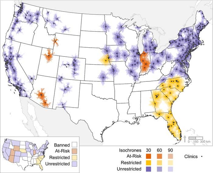
One year after the Dobbs decision, 41.8% of United States women of reproductive age have to drive 30 minutes or more to reach an abortion care facility, according to a study of data as of June 2, 2023.

Researchers predicted that number would rise to 53.5% if other state bills under consideration are passed.

The study estimated longer drives as well, finding that 29.3% of women didn’t have access to a facility within a 60-minute drive and 23.6% lacked access even within a 90-minute drive. Those figures would jump to 45.6% and 43% respectively if new restrictions are passed.

map of continental US shows abortion clinics clustered along costs, some in midwest, and large gaps between across the rest of the country
Locations of physical abortion clinics (black squares), with 30-, 60-, 90-minute driving distance based on current and projected abortion restrictions. (Credit: “A Geospatial Analysis of Abortion Access in the United States after the Reversal of Roe v Wade,” Obstetrics & Gynecology: July 26, 2023)

While the 2022 Dobbs decision overturning Roe v. Wade created a patchwork of abortion restrictions across the nation, the researchers found state laws did not necessarily determine the procedure’s availability.

“This study highlights that abortion access is about more than laws. It’s about more than the state you reside in because people are allowed to cross state borders for medical care. There are a lot of other factors at play,” says Dawn Kopp, the vice chair for OBGYN at Washington State University Elson S. Floyd College of Medicine and senior author of the study in the journal Obstetrics and Gynecology.

For example, Kopp pointed out that in Wisconsin, a state that bans abortion, 61.7% of women still had access within a 90-minute drive to facilities that provide abortions in neighboring states. Conversely, some states where abortion is legal may still not have good access for lack of facilities and the location of facilities relative to where people reside.

For this study, the researchers compared census data for women ages 15-49 to locations of 750 abortion care facilities. The authors note that data limits on gender and age-ranges may not capture all people capable of pregnancy, but it does focus on a large proportion of those affected. The facilities were gathered from lists publicly available online with the intention to mimic what an average person might use if they searched for a provider.

Working with the DataLab at the University of California, Davis, the researchers then used “isochrones,” map lines that create travel time areas using actual roads.

“Use of geospatial technology and analysis in the field of medicine is a growing area of interest,” says coauthor Michele Tobias, a geospatial data specialist at the UC Davis DataLab.

While other studies have estimated distances to care, this research focused on the real drives women would have to make.

“You hear these broad strokes of certain states banning or restricting abortion, but we were able to see on such a granular level the impact this is having on the lives of everyday American women,” says first author Maeve Alterio, a fourth-year medical student at Washington State.

The lack of access has potential serious health consequences as other research has found an increase in maternal and neonatal mortality in states with more abortion restrictions.

These risks have a greater effect on people with lower incomes, the authors said, since the cost of travel and time off work can put the abortion out of reach for people in places like Texas and states without providers in neighboring areas.

Telehealth likely provides access that spans borders for some patients, as many early pregnancy abortions are now done through medication taken at home rather than through a procedure in a clinic. The researchers counted these but could not estimate their geographical reach. They also note the barriers to telehealth including finding follow-up care in areas where abortion is illegal. Many states have also enacted legislation to ban or restrict telehealth use for abortion.

The volatile legal landscape around abortion means the researchers had to re-analyze the data three times as more bills passed or were challenged in court.

Navigating these shifts in the legal landscape has added complexity and uncertainty to the lives of American families, Kopp says.

“This study adds to the stories patients and clinicians have already shared by giving objective data on how pervasive the difficulty to access comprehensive reproductive health care is in a post Roe v. Wade America,” she says.

Source: Washington State University

source

How cells in confinement act like people in crowds

Living cells in confinement behave similarly to people in crowds, adjusting their size while growing alongside other cells in sheets of tissue, researchers report.

John Devany, then a graduate student in the lab of biophysicist Margaret Gardel at the University of Chicago, had been studying epithelial monolayers—sheets of cells that form barriers in skin and coat internal organs—when he noticed something interesting about how the cells were dividing.

“The way people think about division is that a cell will grow to twice its size, divide, and repeat the cycle,” says Devany, the first author of the study in Developmental Cell. But in the epithelial tissue he was observing, division was proceeding as usual, but the daughter cells were ending up smaller than the mother.

The team, collaborating with researchers from New York University, decided to investigate the mechanisms that control cell growth and cycle duration in tissue and discovered that the two processes are not directly coupled.

How cells proliferate—increase in size and number—while in contact with other cells is key to understanding tissue development and growth. A process called “contact inhibition” is thought to restrict cell growth when space becomes limited, controlling tissue overgrowth and preventing tumors, for instance. But the process is regulated by several pathways, and scientists don’t fully understand it.

Gardel, a professor of physics and molecular engineering, studies how living matter emerges from collections of molecules to control the physiology of cells and tissues.

“How cells sense their mechanical environment is critical to stopping them from proliferating,” she says. “And how do the cells actually turn off their biomass production as they feel more constrained? We just don’t know.”

But these results—that the growth pathway is distinct from the cell cycle pathway—help give researchers a framework to study the mechanisms.

Cramped quarters

Many types of cells in our body aren’t packed shoulder to shoulder; they regulate their proliferation through chemical messaging, for instance, or through interactions with their extracellular environment. Epithelial cells form tight barriers, though, able to prevent ions from crossing, and must sense and react to close physical neighbors.

To explore and quantify how tissue growth dynamics regulate cell proliferation, the team used lab-grown layers of mammalian epithelial tissue and observed the cell growth as they went from separate colonies to a layer of merging colonies to tightly packed mature epithelial tissue.

The researchers found that as the tissue expanded and became confined by the size of the culture vessel, cell growth was suppressed, but cell cycle length leading to division was not directly affected. Yet, as the daughter cells became smaller and smaller in volume, they eventually reached a limit at which division did stop.

“The size of organelles, including the nucleus, scale down with the cell’s overall volume,” says Gardel. But the genome size is fixed. “So that’s the limit to how small you can get”—just big enough that the DNA still fits inside.

However, the team found that overexpressing a protein called cyclin D1 allowed the cells to bypass that limit and become even smaller before division halted, indicating that the protein controls the volume-dependent checkpoint. When a cell becomes too small, its genome can get damaged, making those cells particularly prone to becoming cancerous, says Gardel. These findings suggest that cell-volume regulation is key to contact inhibition and the overall health of tissue.

Path towards cancer treatments?

“Most of my lab has worked on questions of cellular scale biophysics,” says Gardel, toward an understanding of the machinery that drives cell behavior. “Our study shows the power of using biophysical and bioengineering approaches to develop new ways to understand tissue scale behaviors.”

“Understanding the mechanisms of growth and division regulation might eventually help develop cancer treatments,” adds Devany. For instance, is the cell volume of small cell cancers—such as some types of lung, prostate, and pancreatic tumors—contributing to new mutations? Maybe that could offer therapeutic targets, Devany suggests.

The dynamics of cells in confinement might also add to the field of tissue engineering, he says, to evolve bioprinting methods and develop material scaffolds that might one day surgically replace damaged tissues. “If we better understand how cells grow and divide in the context of tissue, it might help optimize these kinds of systems.”

How do cells know they’re confined?

Now that Gardel’s team has determined that confined epithelial cells regulate their size and cell cycle separately, one of the next questions is: How do cells know they’re confined? And how do they sense how big they are at any given point?

The researchers were also surprised to find that the cells could communicate over long distances through tissue—some feeling constrained and then telling others to regulate their growth too.

“We think the communication is probably mechanically based,” says Gardel, “but we’re trying to explain the origins of these interactions as well.”

The University of Chicago Functional Genomics processed RNA sequencing samples.

Source: University of Chicago

source

Discovery may treat disease that keeps kids from eating

Researchers have identified a new treatment for EOE, a chronic immune system disease that can prevent children from eating.

Eosinophilic esophagitis (EoE) is triggered by food allergies or airborne allergens which cause a type of white blood cell, eosinophils, to build up in the lining of the esophagus. This causes the esophagus to shorten and the esophageal wall to thicken, making swallowing difficult and causing food to get stuck in the throat.

The disease occurs in an estimated 1 in 2,000 adults but more frequently affects children (1 in 1,500) where symptoms can be harder to diagnose and pose greater risks as difficulty eating can lead to malnutrition, weight loss, and poor growth.

The findings, published in the journal Nature Communications Biology, show the disease is caused by Interleukin-18 (IL-18), a protein involved in the innate immune response that can cause inflammation if produced in excess.

When a food allergen enters the body, it activates a pathway responsible for regulating the innate immune system, resulting in the release of proinflammatory proteins like IL-18. This produces the eosinophils which damage the esophagus.

The researchers discovered that successfully inhibiting this pathway, called the NLRP3 pathway, and the release of IL-18 prevented the development of EoE from both food and airborne allergens.

“Parents and doctors may not be aware of this, but this is a very prominent and serious disease in the pediatric population, and it is increasing in number because it is directly related to food allergens, which are also on the rise,” says lead author Anil Mishra, director of the Eosinophilic Disorder Center at the Tulane University School of Medicine. “In this study, we show that after treating the disease in animals, the disease is gone and completely in remission.”

The findings are crucial for a disease that was not identified until the 1990s. For many years, EoE was misdiagnosed as gastrointestinal reflux disease (GERD), despite GERD medication being ineffective for treating EoE. Additionally, the study’s findings replace decades of thinking that Th2 cells play a major role in triggering EoE.

“Given the paucity of mechanistic information and treatment strategies for EoE, we feel the proposed studies are highly relevant and are poised to have a major impact on establishing the significance of NLRP3-IL-18 pathway in the initiation of EoE pathogenesis,” Mishra says.

The study identifies one existing drug, VX-765, as an inhibitor that may work as a treatment for humans. Importantly, this inhibitor would only deplete pathogenic eosinophils generated and transformed by IL-18 and not affect white blood cells created by IL-5, a protein important for maintaining innate immunity.

Mishra says a clinical trial would be the next step to determining the treatment’s effectiveness.

Source: Tulane University

source