Team finds drug-resistance genes in C. jejuni

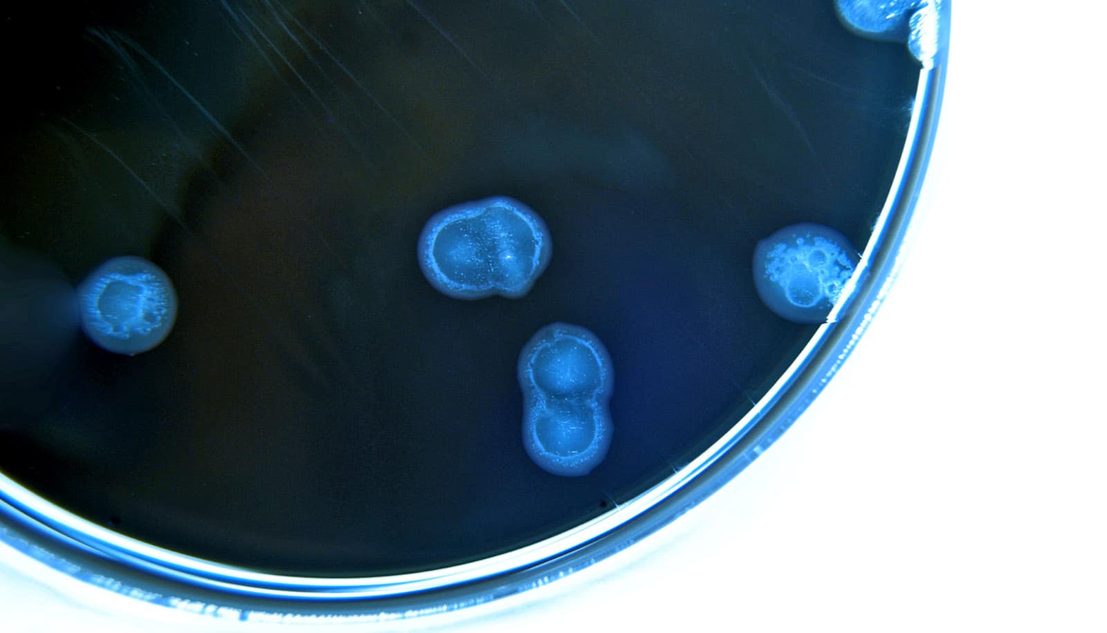
Research finds that antibiotic resistance genes are prevalent in the bacterium Campylobacter jejuni, a leading cause of food borne illness.

The team finds that more than half of the C. jejuni, isolated from patients in Michigan, are genetically protected against at least one antibiotic used to fight bacterial infections. The team’s full report appears in the journal Microbial Genomics.

“We know these pathogens have been around forever but using more sophisticated genome sequencing tools lets us look at them differently,” says Shannon Manning, project leader and a professor in the department of microbiology and molecular genetics at Michigan State University. “We found that the genomes are extremely diverse and contain a lot of genes that can protect them from numerous antibiotics.”

The team’s report provides valuable technical insights to epidemiologists, health care workers, and other specialists, but Manning also emphasizes what the team’s findings mean for the average person.

Although most otherwise healthy adults can fight off such stomach bugs without antibiotics, she says, there are people for whom C. jejuni presents a serious concern. Infections can lead to hospitalization, autoimmune and neurological complications, long-term disability, and even death.

Understanding the extent of antibiotic resistance in this species, as well as which antibiotics different strains are resistant to, can help put patients on better treatment plans sooner.

“If we know the type of antibiotic resistance genes that Campylobacter has, then we know which antibiotics not to give a patient,” Manning says. This can lead to better patient outcomes and shorter hospital stays.

The finding also has broader implications. After people fight off an infection and the pathogen is killed—with or without antibiotics—its genes can linger, including those that provide antibiotic resistance. Other microbes can then pick up those genes, integrate them into their own genomes, and gain resistance.

“That’s really important. Food borne pathogens are ubiquitous. They are found in the foods we eat but also in animals and environments that we come into contact with regularly,” Manning says. “If they carry resistance genes, then not only can they make us sick, but they can also easily transfer the genes to other bacteria.”

This underscores the importance of food hygiene and safety, Manning says, including avoiding cross-contamination of other foods and surfaces before cooking.

The team’s genetic analysis also let the researchers pinpoint the host, or source, of specific strains. That is, they could predict whether the bacteria originated from specific animals or were generalists that are commonly found in multiple hosts.

“When we did this genomic analysis, we found that most patients in Michigan were infected with strains linked to chicken or cattle hosts,” Manning says. Infections also were more likely to occur in rural areas, the team found, suggesting that exposure to these animals and their environments could be important to monitor and potentially control.

Although the Centers for Disease Control and Prevention operates a nationwide network surveilling food borne pathogens, many states, including Michigan, are not part of this system.

“We have unique ecological and agricultural factors in Michigan that may impact how these pathogens survive and proliferate in certain hosts and environments,” says Manning, whose team also studies other major contributors to food borne illness, including E. coli, shigella, and salmonella.

“If you don’t look for them and assess, then you won’t be able to identify which factors are most important for infections and antibiotic resistance or define how Michigan differs from other regions,” she says.

That assessment is, in part, the goal of the Michigan Sequencing Academic Partnership for Public Health Innovation and Response, or MI-SAPPHIRE, a grant that MDHHS awarded Manning’s team last year. The MI-SAPPHIRE program also has support from the CDC.

Source: Matt Davenport for Michigan State University

source

Why you shouldn’t let your guard down when it comes to COVID

More than three years after the COVID pandemic began, we all want to move on. But is it too soon?

With many of us vaccinated at some point, you might be wondering if it’s time to finally let those pandemic worries go for good.

Unfortunately, it’s not so easy, says Angela Branche, an infectious disease physician at the University of Rochester Medicine.

“While we continue to learn more about the virus, which continues to evolve and adapt to evade our immune defense, and figure out new ways to improve protection, our communities should stay alert,” Branche says.

That’s because the likelihood of COVID-19 disappearing any time soon is very low. In fact, scientists expect that the virus will eventually become endemic—causing seasonal illnesses each year.

There is hope that the virus will weaken enough over time, eventually behaving like other coronaviruses—the virus that causes the common cold in most people. However, it’s difficult to tell if or when that will happen.

Just like influenza and other respiratory illnesses, COVID-19 could lead to yearly outbreaks. While protecting yourself from these illnesses does not always mean you will not get sick, you can greatly decrease the intensity of your illness and help protect those around you.

Do I need a booster?

There has been a lot of buzz on boosters lately. They’re like tune-ups for your immune system—helping to extend protection while adding safeguards from new virus variations.

As of August 28, 2023, the Food and Drug Administration and the Centers for Disease Control and Prevention are still determining their official recommendations for COVID-19 boosters during the 2023 fall season.

But, recently the FDA requested development of an updated COVID-19 booster based on newer COVID variants.

In fact, Branche recently led a national study that showed that updated COVID-19 boosters provided better and broader protection against all virus strains (Omicron, Beta, Delta, and Alpha). The results, published in Nature Medicine, definitively showed that boosters no longer need to specifically target the original virus strain to protect against them.

“The newest COVID-19 booster will likely be recommended for those ages 65 and up, as well as those with pre-existing conditions that increase their risk of severe illness,” says Branche.

For healthy adults, whether to get a COVID-19 booster is a risk-based decision that should be made with your primary care provider.

“If you’re heading to college, work in a school or health care institution with frequent contact with many people, it might be best for you to receive a booster,” Branche says. “Remember, receiving a booster will also help reduce severity of infection and possibly the length of illness and transmission to others.”

It’s all about risk versus reward. Getting infected, especially in severe cases, can affect your productivity, cause disruptions in your household and community, and even put vulnerable individuals at risk. The impact is a ripple effect that extends beyond just your own well-being.

‘Every little bit helps’

Remember, vaccines aren’t shields that prevent all infections. They’re designed to prevent severe outcomes and reduce the length of illness. Even if you get infected after getting vaccinated, it’s not a sign that the vaccine didn’t work. It’s all about managing the risk and keeping yourself and your loved ones as safe as possible.

“Every little bit helps. Our goal with vaccinations is to keep everyone safer and healthier,” Branche says.

When it comes to COVID-19, we have come a long way—but that doesn’t mean we should let our guard down. Stay up-to-date on the latest recommendations for boosters and protect yourself and your loved ones to stay as healthy as possible.

Source: University of Rochester

source

Nearly half of dog owners are hesitant to vaccinate their pups

Hesitancy towards COVID vaccines extends to pet vaccinations, as well, and at worrying levels, according to a new study.

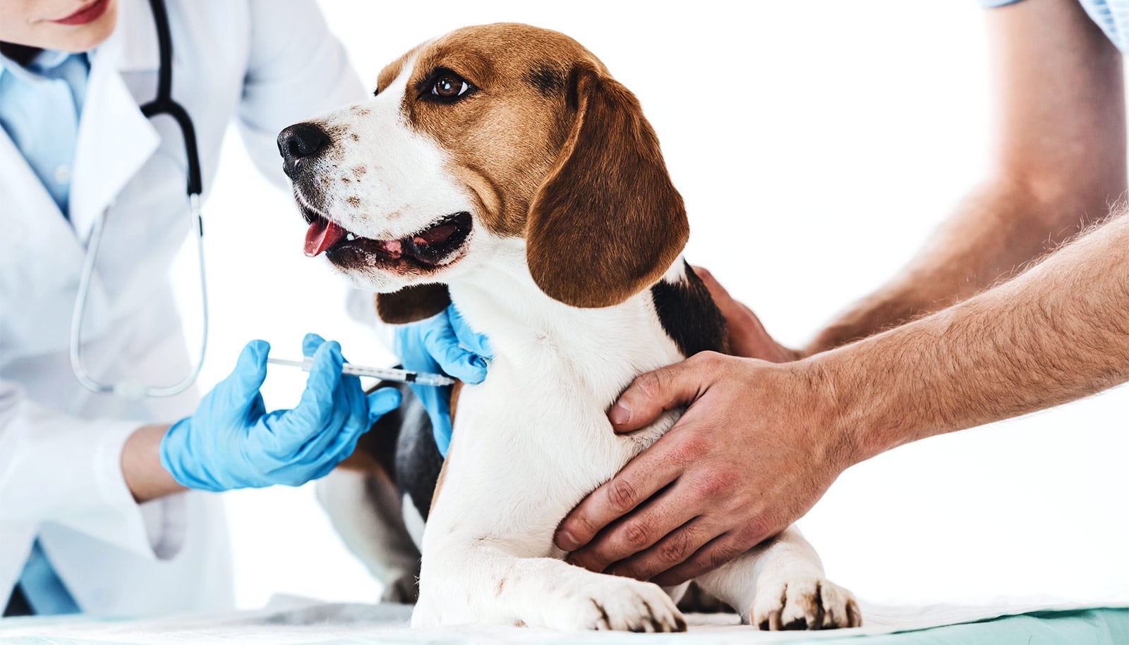
For the study in the journal Vaccine, researchers analyzed a nationally representative sample of adults in the United States and found that more than half of people who own dogs expressed some level of canine vaccine hesitancy—i.e., skepticism about vaccinating their pets against rabies and other diseases.

An estimated 45% of US households own a dog; according to the survey results, nearly 40% of dog owners believe that canine vaccines are unsafe, more than 20% believe these vaccines are ineffective, and 30% consider them to be medically unnecessary.

About 37% of dog owners also believe that canine vaccination could cause their dogs to develop autism, even though there is no scientific data that validates this risk for animals or humans.

The study is the first to formally quantify the prevalence, origins, and health policy consequences of concerns about canine vaccination. Researchers conducted the survey between March 30 and April 10, 2023 among 2,200 dog owners who answered questions through the research sampling firm YouGov.

Notably, the findings show indication of a COVID vaccine “spillover” effect in the US—that people who hold negative attitudes toward human vaccines are more likely to hold negative views toward vaccinating their pets. These dog owners are also more likely to oppose policies that encourage widespread rabies vaccination, and less likely to make the effort to vaccinate their pets.

These attitudes are in contrast to most state-level polices in the US, where almost all states require domestic dogs to be vaccinated against rabies. The disease still poses a potential health threat, as it carries a near 100% fatality rate, and the canine rabies vaccine is much less accessible in developing countries than in the US and other high-income countries. More than 59,000 people die from canine-mediated rabies across the globe each year.

Thus, if fears towards pet vaccinations persist or increase, this skepticism could pose serious public health implications for both animals and humans, the researchers say.

“The vaccine spillover effects that we document in our research underscore the importance of restoring trust in human vaccine safety and efficacy,” says lead and corresponding author Matt Motta, assistant professor of health law, policy, and management at Boston University School of Public Health, who studies how anti-science beliefs and attitudes affect health and health policies. “If non-vaccination were to become more common, our pets, vets, and even our friends and family risk coming into contact with vaccine-preventable diseases.”

The American Animal Hospital Association calls vaccinations “a cornerstone of canine preventive health care” and recommends that all dogs (barring specific medical reasons), receive a core set of vaccines for rabies, distemper, adenovirus, parvovirus, and parainfluenza, and advises that many dogs receive additional “non-core” inoculations for Lyme disease, Bordetella, and other diseases.

Working with animals that are not current on their rabies vaccine poses increased risks for veterinarians and all animal care attendants at a hospital, says coauthor Gabriella Motta, a veterinarian at Glenolden Veterinary Hospital in Glenolden, Pennsylvania. She says she encounters an unvaccinated animal or a vaccine-hesitant pet owner every day in her job.

“When a staff member is bitten by an animal, there is always concern for infection or trauma, but the seriousness of the situation escalates if the animal is unvaccinated or overdue for its rabies vaccine,” Gabriella Motta says.

Per Pennsylvania Department of Agriculture guidelines, once a veterinary staff member is bitten by an undervaccinated or unvaccinated animal, the animal must undergo a period of observation and the staff member is encouraged to receive immediate medical attention. These situations place a mental health burden on the person bitten, as well as the rest of the veterinary staff, in an industry that already struggles with widespread burnout, understaffing, and job turnover, she says.

But she reiterates that the rabies vaccine is overwhelmingly safe and effective.

“With any drug, treatment, or vaccine, there is always a risk of adverse effects, but the risk with the rabies vaccine is quite low—especially when compared to the risk of rabies infection, which is almost 100% lethal,” Gabriella Motta says. Pet owners who are concerned about the cost of pet vaccines may be able to seek low-cost options at local veterinary vaccination clinics, she adds.

The researchers don’t believe canine vaccine hesitancy is widespread enough to pose a current threat to public health in the US, but that could change if vaccine misinformation and mistrust about animal and human vaccines are not quelled with sound, scientific data.

“It’s important to remember that it once seemed unthinkable that MMR [measles, mumps, and rubella] vaccine mandates in public schools might come under attack in state legislatures across the country,” Matt Motta says. “And, yet, previous and ongoing research suggests that this is, indeed, the case.”

Source: Boston University

source

Potato pangenome could lead to a new super spud

Scientists have assembled genome sequences of nearly 300 varieties of potato and its wild relatives to develop a more nutritious, disease-free, and weather-proof crop.

As climate change continues to pose severe challenges to ensuring sustainable food supplies around the world, scientists are looking for ways to improve the resilience and nutritional quality of potatoes.

Martina Strömvik, professor at McGill University, and colleagues have created a potato super pangenome to identify genetic traits that can help produce the next super spud.

“Our super pangenome sheds light on the potato’s genetic diversity and what kinds of genetic traits could potentially be bred into our modern-day crop to make it better,” Strömvik says. “It represents 60 species and is the most extensive collection of genome sequence data for the potato and its relatives to date,” she adds.

A genome is an organism’s complete set of genetic instructions known as the DNA sequence, while a pangenome aims to capture the complete genetic diversity within a species, and a super pangenome also includes multiple species.

The potato is a staple food source for many people around the world—and it’s one of the most important food crops globally, after rice and wheat in terms of human consumption.

Wild potato species can teach us a lot about what genetic traits are critical in adapting to climate change and extreme weather, enhancing nutritional quality, and improving food security,” Strömvik says.

To build the potato pangenome, the researchers used supercomputers to crunch data from public databanks, including gene banks in Canada, the United States, and Peru.

According to the researchers, the pangenome can be used to answer many questions about the evolution of this important crop that was domesticated by Indigenous peoples in the mountains of southern Peru nearly 10,000 years ago. It could also be used to help identify specific genes to create a super spud using traditional breeding or gene editing technology.

“Scientists hope to develop something that can defend against various forms of diseases and better withstand extreme weather like lots of rain, frost, or a drought,” Strömvik says.

The study appears in the Proceedings of the National Academy of Sciences.

Source: Claire Loewen for McGill University

source

Global air pollution is a major health risk

As global pollution edged upward in 2021, so did its burden on human health, according to new data from the Air Quality Life Index released this week.

If the world were to permanently reduce fine particulate pollution (PM2.5) to meet the World Health Organization’s guideline, the average person would add 2.3 years to their life expectancy—or a combined 17.8 billion life-years saved worldwide.

This data makes clear that particulate pollution remains the world’s greatest external risk to human health, with the impact on life expectancy comparable to that of smoking, more than three times that of alcohol use and unsafe water, and more than five times that of transport injuries like car crashes. Yet the pollution challenge worldwide is vastly unequal.

“Three-quarters of air pollution’s impact on global life expectancy occurs in just six countries: Bangladesh, India, Pakistan, China, Nigeria, and Indonesia, where people lose one to more than six years off their lives because of the air they breathe,” says Michael Greenstone, a professor in economics at the University of Chicago and the creator of the AQLI along with colleagues at the Energy Policy Institute at Chicago.

Many polluted places lack basic air pollution infrastructure. Asia and Africa are the two most poignant examples. They contribute 92.7% of life years lost due to pollution. Only 6.8 and 3.7% of governments in Asia and Africa, respectively, provide their citizens with fully open air quality data. Further, just 35.6 and 4.9% of countries in Asia and Africa, respectively, have air quality standards—the most basic building block for policies.

The collective current investments in global air quality infrastructure also do not match where air pollution is having its greatest toll on human life.

While there is a large global fund for HIV/AIDS, malaria, and tuberculosis that annually disburses $4 billion toward the issues, there is no equivalent set of coordinated resources for air pollution. In fact, the entire continent of Africa receives under $300,000 in philanthropic funds toward air pollution. Just $1.4 million goes to Asia, outside of China and India. Europe, the United States, and Canada, meanwhile, receive $34 million, according to the Clean Air Fund.

“Timely, reliable, open air quality data in particular can be the backbone of civil society and government clean air efforts—providing the information that people and governments lack and that allows for more informed policy decisions,” says Christa Hasenkopf, the director of AQLI and air quality programs at EPIC. “Fortunately, we see an immense opportunity to play a role in reversing this by better targeting—and increasing—our funding dollars to collaboratively build the infrastructure that is missing today.”

In no other location on the planet is the deadly impact of pollution more visible than in South Asia, home to the four most polluted countries in the world and nearly a quarter of the global population.

In Bangladesh, India, Nepal, and Pakistan, the AQLI data reveal that residents are expected to lose about five years off their lives on average if the current high levels of pollution persist, and more in the most polluted regions—accounting for more than half of the total life years lost globally due to pollution.

Although the challenge of reducing air pollution around the world may seem daunting, China has had remarkable success, reducing pollution by 42.3% since 2013, the year before the country began a “war against pollution.” Due to these improvements, the average Chinese citizen can expect to live 2.2 years longer, provided the reductions are sustained. However, the pollution in China is still six times higher than the WHO guideline, taking 2.5 years off life expectancy.

Read the full report.

Source: University of Chicago

source

Bat brain ‘GPS’ is also wired for social navigation

The same neurons that help bats navigate through space may also help them navigate collective social environments, according to a new study.

The study appears in the journal Nature.

Many mammals—including bats and humans—are believed to navigate with the help of a brain structure called the hippocampus, which encodes a mental “map” of familiar surroundings. For example, as you walk around your neighborhood or commute to work, individual “place” neurons in the hippocampus fire to indicate where you are.

In the new study, the researchers used wireless neural recording and imaging devices to “listen in” on the hippocampal brain activity of groups of Egyptian fruit bats as they flew freely within a large flight room—often moving among tightly clustered social groups—while tracking technology recorded the bats’ movements.

The researchers were surprised to find that, in this social setting, the bat’s place neurons encoded far more information than simply the animal’s location. As a bat flew toward a landing spot, the firing of place neurons also contained information about the presence or absence of another bat at that spot. And when another bat was present, the activity of these neurons indicated the identity of the bat they were flying toward.

“This is one of the first papers to show identity representation in a non-primate brain,” says study senior author Michael Yartsev, an associate professor of bioengineering and neuroscience at the University of California, Berkeley. “And surprisingly, we found it in the hub of what was supposed to be the brain’s GPS. We found that it still acts as a GPS, but one that is also tuned to the social dynamic in the environment.”

While not as visually stunning as a school of fish or a murmuration of birds, highly social animals like humans and bats also exhibit forms of collective behavior, says first author Angelo Forli, a postdoctoral fellow in Yartsev’s NeuroBat lab.

“Social animals, like humans, will coordinate in space to achieve different goals,” Forli says. “It might be just visiting others. It might be moving together, as in the case of classical collective behaviors or playing a soccer match. Or it might be other forms of cooperation or conflict.”

Due to the complexity of the experiment, Forli initially had doubts about whether allowing groups of bats to fly and interact freely would yield results about the neural basis of collective behavior. He was concerned that the movements of the bats and their social interactions might be too random to uncover robust relationships between their neural activity and their behavior.

So he was pleasantly surprised when the bats spontaneously established a handful of specific resting spots within the flight room and followed very similar trajectories when traveling among them. The bats also showed strong preferences for flying toward specific “friend” bats, often landing very close to or even on top of each other.

“We found that if you put together a small group of bats in a room, they would not actually behave randomly, but would show precise patterns of behavior,” Forli says. “They would spend time with specific individuals and show specific and stable places where they liked to go.”

These precise patterns of behavior allowed Forli to identify not only the neural activity associated with different flight trajectories, but also how the neural activity changed depending on the identity of the bat present at the target location and the movements of other bats.

“By recording just a handful of those neurons from this brain structure, we can really know what the bats were doing in their social space,” Yartsev says. “We could find out if they were going to an empty location or to a location where there were other individuals, which is really surprising.”

In recent years, Yartsev and his NeuroBat Lab have used a variety of wireless neural recording devices and flight tracking technologies to uncover a number of surprising details about the brain, including how bats’ neural activity syncs up while they socialize; how activity in the frontal cortex helps bats identify self vs. others during vocal interactions; how bats’ hippocampus maps not only specific locations, but full flight trajectories; and even how stable spatial memories might be stored in the brain.

This new study brings together the team’s work on navigation and social behavior, showing how these two things are fundamentally intertwined within the brain. The findings also help illuminate why damage to the hippocampus in humans has been linked to both social and spatial aspects of memory loss in neurodegenerative diseases like Alzheimer’s.

“Our episodic memories are a combination of the environment where we are located and our experiences within it—including, of course, our social experiences,” Yartsev says. “Our results are surprising, in the sense that no one has observed this connection before in groups of animals and at the individual neuron level. But they also make sense in that they are very consistent with deficits that people with damage to the hippocampus experience.”

Finally, this study highlights a very important point, Yartsev says. While most of the neuroscientific community examines the brain under “simplified” or “artificial” conditions that are often far removed from the natural behavior the brain has evolved to promote, this work demonstrates the power of the natural approach to neuroscience research.

“For half a century, people have been studying place neurons, but 99% of that work has been done in single animals moving in an empty box,” Yartsev says. “Our findings suggest that there is a lot that can be learned when neuroscience research focuses on natural behavior.”

Support for this research came from the New York Stem Cell Foundation, the National Institute of Mental Health, the Air Force Office of Scientific Research, the Packard Fellowship, the National Institute of Neurological Disorders and Stroke, the Vallee Foundation, the Office of Naval Research, the Searle Scholars Program, the Human Frontiers Fellowship, and the European Molecular Biology Organization.

Source: UC Berkeley

source

How hogfish ‘see’ with their skin

Hogfish don’t just see with their eyes. They also see with their skin—and now researchers think they know why.

A few years ago while on a fishing trip in the Florida Keys, biologist Lori Schweikert came face to face with an unusual quick-change act. She reeled in a pointy-snouted reef fish called a hogfish and threw it onboard. But later when she went to put it in a cooler she noticed something odd: its skin had taken on the same color and pattern as the deck of the boat.

A common fish in the western Atlantic Ocean from North Carolina to Brazil, the hogfish is known for its color-changing skin. The species can morph from white to mottled to reddish-brown in a matter of milliseconds to blend in with corals, sand, or rocks.

Still, Schweikert was surprised because this hogfish had continued its camouflage even though it was no longer alive. Which got her wondering: can hogfish detect light using only their skin, independently of their eyes and brain?

“That opened up this whole field for me,” Schweikert says.

In the years that followed, Schweikert started researching the physiology of “skin vision” as a postdoctoral fellow at Duke University and Florida International University.

In 2018, Schweikert and Duke biologist Sönke Johnsen published a study showing that hogfish carry a gene for a light-sensitive protein called opsin that is activated in their skin, and that this gene is different from the opsin genes found in their eyes.

Other color-changing animals from octopuses to geckos have been found to make light-sensing opsins in their skin, too. But exactly how they use them to help change color is unclear.

“When we found it in hogfish, I looked at Sönke and said: Why have a light detector in the skin?” says Schweikert, now an assistant professor at the University of North Carolina, Wilmington.

One hypothesis is that light-sensing skin helps animals take in their surroundings. But new findings suggest another possibility—”that they could be using it to view themselves,” Schweikert says.

In a study in the journal Nature Communications, Schweikert, Johnsen, and colleagues teamed up to take a closer look at hogfish skin.

The researchers took pieces of skin from different parts of the fish’s body and took pictures of them under a microscope.

Up close, a hogfish’s skin looks like a pointillist painting. Each dot of color is a specialized cell called a chromatophore containing granules of pigment that can be red, yellow, or black.

It’s the movement of these pigment granules that changes the skin color. When the granules spread out across the cell, the color appears darker. When they cluster together into a tiny spot that’s hard to see, the cell becomes more transparent.

Next, the researchers used a technique called immunolabeling to locate the opsin proteins within the skin. They found that in the hogfish, opsins aren’t produced in the color-changing chromatophore cells. Instead, the opsins reside in other cells directly beneath them.

Images taken with a transmission electron microscope revealed a previously unknown cell type, just below the chromatophores, packed with opsin protein.

This means that light striking the skin must pass through the pigment-filled chromatophores first before it reaches the light-sensitive layer, Schweikert says.

The researchers estimate that the opsin molecules in hogfish skin are most sensitive to blue light. This happens to be the wavelength of light that the pigment granules in the fish’s chromatophores absorb best.

The findings suggest that fish’s light-sensitive opsins act somewhat like internal Polaroid film, capturing changes in the light that is able to filter through the pigment-filled cells above as the pigment granules bunch up or fan out.

“The animals can literally take a photo of their own skin from the inside,” Johnsen says. “In a way they can tell the animal what it’s skin looks like, since it can’t really bend over to look.”

“Just to be clear, we’re not arguing that hogfish skin functions like an eye,” Schweikert adds. Eyes do more than merely detect light—they form images. “We don’t have any evidence to suggest that’s what’s happening in their skin,” Schweikert says.

Rather, it’s a sensory feedback mechanism that lets the hogfish monitor its own skin as it changes color, and fine-tune it to fit what it sees with its eyes.

“They appear to be watching their own color change,” Schweikert says.

The researchers say the work is important because it could pave the way to new sensory feedback techniques for devices such as robotic limbs and self-driving cars that must fine-tune their performance without relying solely on eyesight or camera feeds.

“Sensory feedback is one of the tricks that technology is still trying to figure out,” Johnsen says. “This study is a nice dissection of a new sensory feedback system.”

“If you didn’t have a mirror, and you couldn’t bend your neck, how would you know if you’re dressed appropriately?” Schweikert says. “For us it may not matter,” she adds. But for creatures that use their color-changing abilities to hide from predators, warn rivals or woo mates, “it could be life or death.”

Additional researchers from the Florida Institute of Technology, Florida International University, and the Air Force Research Laboratory contributed to the work. Financial support came from Duke University, Florida International University, the Marine Biological Laboratory, and the National Science Foundation.

Source: Duke University

source

Employers judge mental health posts on social media

Mental health disclosures on platforms like LinkedIn can affect the way potential employers view job applicants, a study finds.

“People are often encouraged to discuss their mental health struggles on social media with the goal of reducing the stigma associated with mental health challenges,” says Lori Foster, coauthor of a paper on the study and a professor of psychology at North Carolina State University.

“We think reducing stigma around mental health is extremely important, but our study suggests that mental health posts on platforms such as LinkedIn could have unforeseen consequences for people disclosing their mental health challenges.”

“Specifically, we found that these disclosures can influence the way people view us in professional contexts,” says Jenna McChesney, first author of the study and assistant professor of psychology at Meredith College who worked on the study while a grad student at NC State. “It’s important for people to take that into consideration when determining whether to share their mental health experiences online.”

For the study, the researchers set out to determine the extent to which posts about mental health on LinkedIn affected perceptions of an individual’s personality and future performance in the workplace.

To address the question, the researchers enlisted 409 professionals with hiring experience to participate in a study. About 25% of the participants saw the LinkedIn page of a job candidate, with no mention of mental health challenges. Another 25% of the participants saw the same LinkedIn page, but it included a post mentioning the candidate’s experiences with anxiety and depression. A third quarter of the study participants saw the LinkedIn profile and heard an audio interview with the candidate. And the last 25% of participants saw the LinkedIn profile, including the post about anxiety and depression, and heard the audio interview. All of the study participants were then asked a series of questions about the job candidate’s personality and future performance in the workplace.

“We found that study participants who saw the LinkedIn post about mental health challenges viewed the job candidate as being less emotionally stable and less conscientious,” McChesney says. “Hearing the interview lessened a study participant’s questions about the candidate’s emotional stability, but only slightly. And hearing the interview did not affect the views of participants about the job candidate’s conscientiousness. In other words, the perceptions evaluators had after seeing the LinkedIn profile largely persisted throughout the interview.

“Our findings don’t mean people should refrain from posting about anxiety and depression on LinkedIn,” McChesney says. “However, people who are considering posting about these issues should be aware that doing so could change future employers’ perceptions of them.”

“There is a big push for people to always be their full authentic selves, but there has been little research into any positive or negative consequences associated with that,” Foster says. “This study is a step toward getting a more complete picture, and it highlights just how much additional work is needed.

“There are also implications for employers,” Foster says. “When hiring managers look up candidates on LinkedIn, they risk seeing information that can color their perceptions, even subconsciously. Organizations should implement guidelines for using LinkedIn during the hiring process to encourage equitable comparisons among all candidates, including those who openly discuss mental health challenges.”

The paper appears in the Journal of Business and Psychology.

Source: NC State

source

Blood test could detect Parkinson’s sooner

A new blood test that detects Parkinson’s disease, potentially establishing a way to diagnose the condition before nervous system damage worsens.

The new blood-based diagnostic test would be a major advancement for Parkinson’s disease, which afflicts 10 million people worldwide and is the second-most common neurodegenerative disease after Alzheimer’s.

“Currently, Parkinson’s disease is diagnosed largely based on clinical symptoms after significant neurological damage has already occurred,” says senior study author Laurie Sanders, an associate professor in the Duke University School of Medicine’s departments of neurology and pathology and member of the Duke Center for Neurodegeneration and Neurotherapeutics.

“A simple blood test would allow us to diagnose the disease earlier and start therapies sooner,” Sanders says. “Additionally, a clear-cut diagnosis would accurately identify patients who could participate in drug studies, leading to the development of better treatments and potentially even cures.”

As a biomarker for their diagnostic tool, Sanders and colleagues focused on DNA damage in the mitochondria. Mitochondria are factories within cells that convert raw energy into a form that powers cells. They contain their own DNA, which can undergo damage separately from the nuclear DNA that encodes most of an organism’s genome.

Earlier studies have associated mitochondrial DNA damage with an increased risk of Parkinson’s disease, and the researchers had previously reported an accumulation of mitochondrial DNA damage specifically in the brain tissue of deceased Parkinson’s patients.

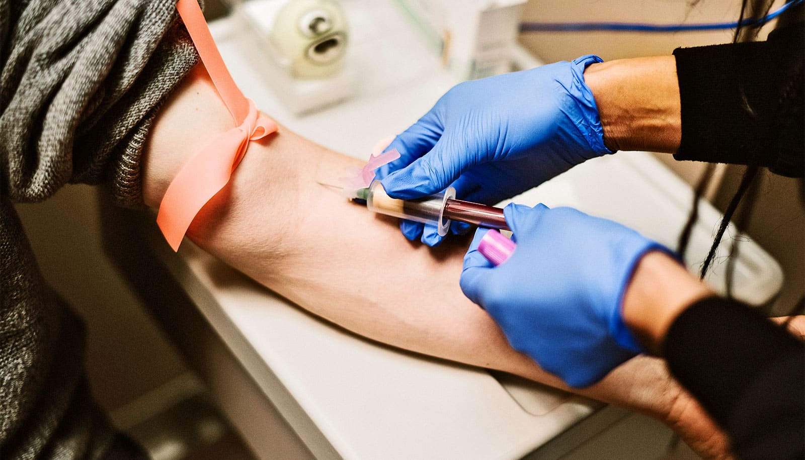
Using polymerase chain reaction (PCR) technology, the researchers developed an assay that successfully quantified higher levels of mitochondrial DNA damage in blood cells collected from patients with Parkinson’s disease compared to people without the disease.

The new test also identified high levels of the damaged DNA in the blood samples of people who harbor the genetic mutation LRRK2, which has been associated with an increased risk of the disease. The assay was able to detect Parkinson’s disease patients with and without LRRK2 mutations.

A further analysis in cells from patients with Parkinson’s disease explored whether the team’s PCR-based test could determine the impact of a therapy targeting the effects associated with LRRK2 mutation.

In these samples, the test identified lower mitochondrial DNA damage in cells treated with a LRRK2 inhibitor compared to samples from patients who did not receive the inhibitor. This suggests the assay could help pinpoint Parkinson’s disease patients who might benefit from LRRK2 kinase inhibitor treatments, even if they do not have the LRRK2 mutation.

“Our hope is that this assay could not only diagnose Parkinson’s disease, but also identify drugs that reverse or halt mitochondrial DNA damage and the disease process,” Sanders says. “This disease takes a terrible toll on people, and we are still just treating the symptoms. It’s important to get new, effective treatments over the finish line.”

The researchers’ future will include further testing of the assay in samples from patients with the earliest stages of disease, before symptoms develop.

The study, published in Science Translational Medicine, received support in part from the Michael J. Fox Foundation for Parkinson’s Research; the Mitochondria, Aging & Metabolism Seed Grant Program; the William N. & Bernice E. Bumpus Foundation; the Pepper Center at the University of Pittsburgh; the Medical Research Council; the Chief Scientist Office Senior Clinical Academic Fellowship; the National Health and Medical Research Council of Australia Leadership Fellowship; Busch Biomedical Research; and the National Institutes of Health.

Source: Duke University

source

Early in pandemic, half a million kids moved in with grandparents

Grandparents appear to have served as an important private safety net when COVID-19 first hit the US, according to a new study.

The findings show that the pandemic’s arrival in 2020 coincided with a surge of nearly 510,000 children living in “doubled-up” households, co-residing with other adults in addition to their parents or parents’ partners.

While these living arrangements had already been increasing before COVID-19, this was an additional increase beyond what would be expected based on previous trends—and most of those children, about 460,000, moved in with grandparents.

The surge was temporary, however—those households returned to expected levels in 2021, according to the study in the journal Demography.

“Despite the health risks associated with co-residing with larger families during COVID, economic and instrumental needs still led Americans to live together. I think that really speaks to the strength and importance of family ties as a safety net in the US,” says lead author Mariana Amorim, a sociologist at Washington State University.

Using survey data collected by the US Census, Amorim and coauthor Natasha Pilkauskas of the University of Michigan analyzed the trends for children in doubled-up households from 2015-2021.

About 15.3% of children lived in these types of households in 2015, and there was an incremental average increase of about 0.1 percentage points each year, but from 2019 to 2020 the share of children in these arrangements jumped from 15.7% to 16.3%, an increase six times greater than expected.

The people moving into these doubled-up arrangements tended to be families headed by single mothers and mothers who had never married or who were not working as well as families with children under the age of six. There was also a larger than usual increase in Black and Hispanic children living in multigenerational households.

These demographics suggest the surge in co-residence was driven by both the need for economic and other instrumental support–such as childcare and perhaps to some degree elder care on the part of the grandparents, the researchers say. These needs appeared to outweigh concerns over spreading COVID-19 to more vulnerable older relatives.

The researchers also uncovered a seasonality in trends of “doubling up.” Before the pandemic, increases in these co-residing households tended to follow a pattern with more co-residence occurring in late fall and winter. The researchers speculate that this may follow already established seasonal patterns of births and divorces.

When the pandemic hit, it upended that seasonal pattern, with more people moving into doubled-up households in spring and summer of 2020 before decreasing later in that year and normalizing in 2021.

“We found this type of co-residence to be really short lived. This really drives home the idea that doubling up, particularly living with grandparents, is a strategy that’s used to address economic or instrumental needs, but it’s not really the preference for Americans,” says Amorim. “As soon as people are able to move out and live independently, they do.”

Source: Washington State University

source