Team sees light make atoms ‘dance’ in perovskites

New research shows how the atoms in perovskites move in response to light.

The breakthrough in visualization supports the researchers’ efforts to squeeze every possible drop of utility out of perovskite-based materials, including solar cells, a long-standing project that only recently yielded an advance to make the devices far more durable.

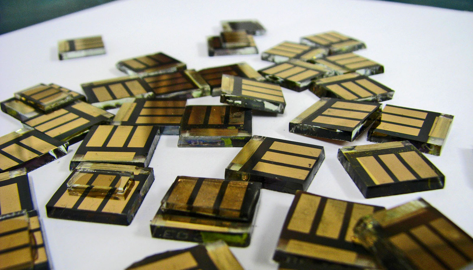
The study in Nature Physics details the first direct measurement of structural dynamics under light-induced excitation in 2D perovskites. Perovskites are layered materials that have well-ordered crystal lattices. They are highly efficient harvesters of light that are being explored for use as solar cells, photodetectors, photocatalysts, light-emitting diodes, quantum emitters, and more.

“The next frontier in light-to-energy conversion devices is harvesting hot carriers,” Aditya Mohite, a corresponding author of the study. “Studies have shown that hot carriers in perovskite can live up to 10-100 times longer than in classical semiconductors. However, the mechanisms and design principles for the energy transfer and how they interact with the lattice are not understood.”

Hot carriers are short-lived, high-energy charge carriers, either electrons for negative charges or electron “holes” for positive charges, and having the ability to harvest their energy would allow light-harvesting devices to “surpass thermodynamic efficiency,” says Mohite, an associate professor of chemical and biomolecular engineering in Rice University’s George R. Brown School of Engineering.

Mohite and three members of his research group, senior scientist Jean-Christophe Blancon and graduate students Hao Zhang and Wenbin Li, worked with colleagues at the SLAC National Accelerator Laboratory to see how atoms in a perovskite lattice rearranged themselves when a hot carrier was created in their midst. They visualized lattice reorganization in real time using ultrafast electron diffraction.

“Whenever you expose these soft semiconductors to stimuli like electric fields, interesting things happen,” Mohite says. “When you generate electrons and holes, they tend to couple to the lattice in unusual and really strong ways, which is not the case for classical materials and semiconductors.

“So there was a fundamental physics question,” he says. “Can we visualize these interactions? Can we see how the structure is actually responding at very fast timescales as you put light onto this material?”

The answer was yes, but only with a strong input. SLAC’s mega-electron-volt ultrafast electron diffraction (MeV-UED) facility is one of the few places in the world with pulsed lasers capable of creating the electron-hole plasma in perovskites that was needed to reveal how the lattice structure changed in less than a billionth of a second in response to a hot carrier.

“The way this experiment works is that you shoot a laser through the material and then you send an electron beam that goes past it at a very short time delay,” Mohite explains. “You start to see exactly what you would in a TEM (transmission electron microscope) image. With the high-energy electrons at SLAC, you can see diffraction patterns from thicker samples, and that allows you to monitor what happens to those electrons and holes and how they interact with the lattice.”

The experiments at SLAC produced before-and-after diffraction patterns that Mohite’s team interpreted to show how the lattice changed. They found that after the lattice was excited by light, it relaxed and literally straightened up in as little as one picosecond, or one-trillionth of a second.

Zhang says, “There’s a subtle tilting of the perovskite octahedra, which triggers this transient lattice reorganization towards a higher symmetric phase.”

By demonstrating that a perovskite lattice can suddenly become less distorted in response to light, the research showed it should be possible to tune how perovskite lattices interact with light, and it suggested a way to accomplish the tuning.

Li says, “This effect is very dependent on the type of structure and type of organic spacer cation.”

There are many recipes for making perovskites, but all contain organic cations, an ingredient that acts as a spacer between the materials’ semiconducting layers. By substituting or subtly changing organic cations, researchers could tailor lattice rigidity, dialing it up or down to alter how the material responds to light, Li says.

Mohite says the experiments also show that tuning a perovskite’s lattice alters its heat-transfer properties.

“What is generally expected is that when you excite electrons at a very high energy level, they lose their energy to the lattice,” he says. “Some of that energy is converted to whatever process you want, but a lot of it is lost as heat, which shows in the diffraction pattern as a loss in intensity.

“The lattice is getting more energy from thermal energy,” Mohite says. “That’s the classical effect, which is expected, and is well-known as the Debye-Waller factor. But because we can now know exactly what’s happening in every direction of the crystal lattice, we see the lattice starts to get more crystalline or ordered. And that’s totally counterintuitive.”

A better understanding of how excited perovskites handle heat is a bonus of the research, he says.

“As we make devices smaller and smaller, one of the biggest challenges from a microelectronics perspective is heat management,” Mohite says. “Understanding this heat generation and how it’s being transported through materials is important.

“When people talk about stacking devices, they need to be able to extract heat very fast,” he says. “As we move to new technologies that consume less power and generate less heat, these types of measurements will allow us to directly probe how heat is flowing.”

The research had support from the Department of Energy, the Office of Naval Research, the Robert A. Welch Foundation, and the Academic Institute of France.

Source: Rice University

source

People who share ideology have similar ‘neural fingerprints’

People who share a political ideology have more similar “neural fingerprints” of political words and process new information in similar ways, according to a new analysis.

Take the word “freedom,” for example, or a picture of the American flag, or even the 2020 US presidential election. A person who identifies politically as liberal vs. one who identifies as conservative will likely have opposing interpretations when processing this information—and the new research helps to explain why.

While previous theories posited that political polarization results from selective consumption (and over-consumption) of news and social media, a team led by researchers at Brown University hypothesized that polarization may start even earlier.

The new study appears in Science Advances.

Individuals who share an ideology have more similar neural fingerprints of political words, experience greater neural synchrony when engaging with political content, and their brains sequentially segment new information into the same units of meaning.

In this way, the researchers say, they show how polarization arises at the very point when the brain receives and processes new information.

“This research helps shed light on what happens in the brain that gives rise to political polarization,” says senior study author Oriel FeldmanHall, an associate professor of cognitive, linguistic, and psychological sciences who is affiliated with the Carney Institute of Brain Science at Brown University. Daantje de Bruin, a graduate student in FeldmanHall’s lab, led the research and conducted the data analysis.

Previous research from FeldmanHall’s lab showed that when watching a potentially polarizing video about hot-button issues like abortion, policing, or immigration, the brain activity of people who identified as Democrat or Republican was similar to the brain activity of people in their respective parties.

That neurosynchrony, FeldmanHall explains, is considered evidence that the brains are processing the information in a similar way. For this new study, the researchers wanted to get an even more detailed picture of why and how the brains of people in the same political party are able to sync up.

To do that, the team used a range of methods that they say have never before been used in conjunction with each other. They conducted a series of experiments with a group of 44 participants, equally split among liberals and conservatives, who agreed to perform various cognitive tasks while undergoing functional magnetic resonance imaging (fMRI), which measures the small changes in blood flow that occur with brain activity.

This research helps shed light on what happens in the brain that gives rise to political polarization.

Participants first completed a word reading task in which they were presented with single words (e.g., “immigration,” “abortion”) and asked to determine whether the word was political or non-political (indicated via a button press). Then the participants watched a series of videos, including a neutrally worded news clip on abortion and a heated 2016 vice presidential campaign debate on police brutality and immigration. During the experiments, the participants’ brain activity was measured using fMRI.

One of the methods the researchers used is called representation similarity analysis. When a person sees a simple, static image, like a word, the brain will represent that word with certain activity patterns.

“You can think of it as the brain representing the word by firing neurons in a certain way,” FeldmanHall says. “It’s almost like a fingerprint—a neural fingerprint that encodes the concept of that word within the brain.”

She added that since neural activity patterns store information about the world, how the brain represents this information is considered a metric for how that information is interpreted and used to steer behavior and attitudes.

In the study, the participants were exposed to words that are often politicized, like “abortion,” “immigration” and “gangs,” as well as more ambiguous words, like “freedom”.

The researchers found by analyzing the fMRI data that the neural fingerprint created by a liberal brain is more similar to other liberal brains than the neural fingerprint created by a conservative brain, and vice versa. This is important, FeldmanHall says, because it shows how the brains of partisans are processing information in a polarized way, even when it’s devoid of any political context.

The researchers also used a newer methodology called neural segmentation to explore how the brains of people who identify with a particular party bias the interpretation of incoming information. Brains are constantly receiving visual and auditory input, FeldmanHall says, and the way the brain makes sense of that continuous barrage of information is to separate it into discrete chunks, or segments.

“It’s like dividing a book of solid text into sentences, paragraphs, and chapters,” she says.

The researchers found that the brains of Democrats separate incoming information in the same way, which then gives similar, partisan meanings to those pieces of information—but that the brains of Republicans segment the same information in a different way.

The researchers note that individuals who shared an ideology had more similar neural representations of political words and experienced greater neural synchrony while watching the political videos, and segmented real-world information into the same meaningful units.

“The reason two liberal brains are synchronizing when watching a complicated video is due in part to the fact that each brain has neural fingerprints for political concepts or words that are very aligned,” FeldmanHall explains.

This explains why two opposing partisans can watch the same news segment and both believe that it was biased against their side—for each partisan, the words, images, sounds, and concepts were represented in their brain in a different way (but similar to other partisans who share their ideology). The stream of information was also segmented out in a different format, telling a different ideological story.

Taken together, the researchers conclude, the findings show that political ideology is shaped by semantic representations of political concepts processed in an environment free of any polarizing agenda, and that these representations bias how real-world political information is construed into a polarized perspective.

“In this way, our study provided a mechanistic account for why political polarization arises,” FeldmanHall says.

The researchers are now focusing on how this explanation of polarization can be used to combat polarization.

“The problem of political polarization can’t be addressed on a superficial level,” FeldmanHall says. “Our work showed that these polarized beliefs are very entrenched, and go all the way down to the way people experience a political word. Understanding this will influence how researchers think about potential interventions.”

Additional contributors to this research include Pedro L. Rodríguez from the Center for Data Science at New York University and Jeroen M. van Baar from the Netherlands Institute of Mental Health and Addiction.

Source: Brown University

source

Pee is a big part of giraffe sex lives

New research provides insight into the unique sex lives of giraffes, their reproductive behavior, and how their anatomy supports that behavior.

It can be hard to know if someone is really into you. Sometimes, you get hints—a certain look or smile, a nervous blush, or flirtation. Giraffes get none of that.

They have no set breeding season. They don’t go into heat, like dogs or cats. They don’t make mating calls or provide visual cues of sexual readiness. So how is a male giraffe to know his advances will be well-received? In short: pee, pheromones, and a gentle nudge.

The new study in the journal Animals describes how male giraffes test females for sexual receptivity.

First, the bulls provoke the females to urinate by nudging them and sniffing their genitalia. If the female is open to his invitation, she widens her stance and pees for about 5 seconds while the male takes the urine in his mouth. He then curls his lip, inhaling with an open mouth—an act called flehmen that transports the female’s scent and pheromones from his oral cavity to the vomeronasal organ.

The study provides the most precise understanding yet of how flehmen occurs with giraffes’ anatomy. While flehmen is common among many animals, including horses and cats, most mammals wait until urine is on the ground to investigate. The giraffe, however, is not built for such explorations.

“They don’t risk going all the way to the ground because of the extreme development of their head and neck,” says lead author Lynette Hart, a professor of population health and reproduction in the School of Veterinary Medicine at the University of California, Davis. “So they have to nudge the female, effectively saying, ‘Please urinate now.’ And often she will. He has to elicit her cooperation. If not, he’ll know there’s no future for him with her.”

Hart and her coauthor and husband, Benjamin Hart, professor emeritus with the School of Veterinary Medicine, witnessed this behavior on multiple research trips to Namibia’s Etosha National Park.

Dotted among the park’s western side were large watering holes, where dozens of giraffes would congregate. Lynette called it “a dream come true” for observing giraffes. “So often you see a few in the distance, not an up-close view of what they’re doing,” she says.

Benjamin Hart had studied how flehmen behavior worked within the anatomy of other animals, including goats. During their trips to East Africa, the Harts suspected a similar process was underway for giraffes.

“This is part of their reproductive behavior,” Benjamin Hart says. “This adds to our understanding of what giraffes are doing as they accumulate around a water hole. People love watching giraffes. I think the more the public understands about them, the more interested they’ll be in their conservation.”

The Harts also describe in the study previously undocumented giraffe behaviors, from chewing bones to potentially mourning their dead:

  • Earlier studies noted that osteophagia, or chewing bones, was unusual for giraffes. But the Harts observed many instances of giraffes seeking and chewing bones, and sometimes getting them lodged in their mouths.
  • After a giraffe had been killed by two lions, the Harts also witnessed for several days a steady procession of giraffes arriving to investigate the body.
  • The Harts experienced another significant observation when they heard a bull emit a loud growl on different occasions. It was most likely a warning call, as it drove away most surrounding giraffes. Giraffes are typically very quiet and were once even thought to be mute.

The research received no direct external funding. Financial assistance for travel and accommodations was provided by UC Berkeley’s University Research Expeditions Program.

Source: UC Davis

source

New childhood obesity guidelines may do more harm than good

New guidelines from the American Academy of Pediatrics on how to deal with childhood obesity will have “unintended negative effects,” says Kate Bauer.

The guidelines, the first in 15 years, advise urgent and early treatment interventions, including medications and surgery at younger ages, rather than relying on wait-and-see treatments.

More than 14.4 million US children and teens are at risk of serious short and long-term health concerns such as cardiovascular disease, diabetes, and behavioral health issues, if untreated, according to the AAP.

Bauer, an associate professor of nutritional sciences at the University of Michigan School of Public Health, is an expert on causes of and reactions to childhood obesity, especially in marginalized communities.

Here, she talks about the guidelines and some of her specific concerns about what they mean for health care providers, families, and society:

Source: Kim North Shine for University of Michigan

source

To spare research volunteers, robot gets mosquito bites

A new system for tropical disease research spares people and animals mosquito bites.

Researchers are working to take some of the pain out of studying the feeding behavior of mosquitoes. The insects’ bites can spread diseases like malaria, dengue, and yellow fever, but setting up experiments to examine their behavior can take a big bite out of lab budgets.

“Many mosquito experiments still rely on human volunteers and animal subjects,” says Kevin Janson, a graduate student in bioengineering at Rice University and lead coauthor of a study in Frontiers in Bioengineering and Biotechnology. Live subject testing can be expensive, and Janson says the “data can take many hours to process.”

So he and his coauthors found a way to automate the collection and processing of that data using inexpensive cameras and machine-learning software. To eliminate the need for live volunteers, their system uses patches of synthetic skin made with a 3D printer. Each patch of gelatin-like hydrogel comes complete with tiny passageways that can be filled with flowing blood.

To create the stand-ins for skin, the team used bioprinting techniques that were pioneered in the lab of former Rice professor Jordan Miller.

For feeding tests, as many as six of the hydrogels can be placed in a transparent plastic box about the size of a volleyball. The chambers are surrounded with cameras that point at each blood-infused hydrogel patch. Mosquitos go in the chamber, and the cameras record how often they land at each location, how long they stay, whether or not they bite, how long they feed, etc.

Researchers in the laboratory of Dawn Wesson, associate professor of tropical medicine at Tulane University, tested the system. Wesson’s research group has facilities for breeding and testing large populations of mosquitoes of varying species.

In the proof-of-concept experiments featured in the study, Wesson, Janson, and coauthors used the system to examine the effectiveness of existing mosquito repellents made with either DEET or a plant-based repellent derived from the oil of lemon eucalyptus plants. Tests showed mosquitoes readily fed on hydrogels without any repellent and stayed away from hydrogel patches coated with either repellent. While DEET was slightly more effective, both tests showed each repellent deterred mosquitoes from feeding.

Omid Veiseh, the study’s corresponding author and an assistant professor of bioengineering at Rice, says the results suggest the behavioral test system can be scaled up to test or discover new repellents and to study mosquito behavior more broadly. He says the system also could open the door for testing in labs that couldn’t previously afford it.

“It provides a consistent and controlled method of observation,” Veiseh says. “The hope is researchers will be able to use that to identify ways to prevent the spread of disease in the future.”

Wesson says her lab is already using the system to study viral transmission of dengue, and she plans to use it in future studies involving malaria parasites.

“We are using the system to examine virus transmission during blood feeding,” Wesson says. “We are interested both in how viruses get taken up by uninfected mosquitoes and how viruses get deposited, along with saliva, by infected mosquitoes.

“If we had a better understanding of the fine mechanics and proteins and other molecules that are involved, we might be able to develop some means of interfering in those processes,” she says.

The Robert J. Kleberg, Jr. and Helen C. Kleberg Foundation supported the research.

Source: Rice University

source

Abortion restrictions may stymie miscarriage care

People experiencing a miscarriage in states with restrictive abortion policies may be less likely to receive optimal care than those in states with supportive abortion policies.

The new study, published in the journal Women’s Health Issues, was conducted prior to the Supreme Court’s decision last June to overturn Roe v. Wade when lead author Elana Tal was a fellow at Washington University School of Medicine in St. Louis.

Tal, now a clinical assistant professor in the obstetrics and gynecology department in the Jacobs School of Medicine and Biomedical Sciences at the University at Buffalo, had concerns about how restrictive abortion policies affect care for people experiencing spontaneous pregnancy loss.

“I had a hunch that restricting abortion means less ideal care for people experiencing miscarriage,” says Tal, who focuses on complex family planning.

“Too often, when we talk about abortion the conversation becomes about the morality of ending a pregnancy and not about how restricting abortion affects reproductive health in general,” she says. “We know abortion restrictions correlate with higher rates of maternal mortality, so it follows that other aspects of health care would be affected, especially miscarriage care, which so closely mirrors abortion care. I wanted to find out if that was true.”

Spontaneous pregnancy loss, i.e. miscarriage, in the first trimester occurs in about 10% of all clinically recognized pregnancies, and 25% of all people capable of becoming pregnant will experience a miscarriage in their lifetime.

This study is among the first to explore how miscarriages are managed in light of evidence-based, patient-centered guidelines issued in recent years by the American College of Obstetricians and Gynecologists (ACOG).

Those guidelines, and the research they were based on, found that for managing early pregnancy loss, optimal care includes uterine aspiration in the physician’s office and the prescribing of both mifepristone and misoprostol, which block hormones that are necessary for pregnancy and help clear the uterus.

Because these methods are also used to terminate a pregnancy, Tal and her coauthors wanted to see if access to these methods is compromised for those experiencing a miscarriage.

The researchers found that in states with restrictive abortion policies, physicians managing early pregnancy loss were less likely than physicians in supportive states (40.8% vs. 67.5%) to offer mifepristone alone and less likely to offer both mifepristone and office uterine aspiration (33.2% vs. 51.3%). They also found, however, that there was no significant difference in the proportion offering uterine aspiration between physicians in restrictive states and those in supportive states.

In addition, physicians in restrictive states were less likely to report having received abortion training (67.3% vs. 89.6%), and less likely to report perceived institutional support for abortion care (49% vs. 85%).

“Our study is consistent with the notion that general pregnancy care suffers where abortion is restricted,” says Tal. “That would apply to routine early miscarriage, more complicated miscarriage like second trimester fetal demise, and abortion for life-threatening situations.

“Clinicians should be aware of the potential deficiencies in their ability to provide miscarriage care if they train or practice in states with restrictive laws,” she says.

In addition to surveying the impact of a state’s policies on access to reproductive care, the survey was also aimed at determining how a physician’s perception of their institution’s support for abortion care, or lack of it, might influence access to reproductive care generally.

The survey was sent to more than 1,500 members of ACOG. Respondents were deemed eligible to respond if they were an attending physician with an academic medical center who provided obstetric and/or gynecologic care and had provided early pregnancy loss care in the past year.

Eligible responses were received from 350 physicians from every region in the US, representing half of academic medicine centers.

Tal notes that the end of Roe v. Wade is expected to even more strongly affect access to care for those experiencing miscarriage.

“At the time of our study, access to abortion was constitutionally protected, and we still saw disparities in the management of miscarriage, a very common reproductive health issue,” she says. “We would expect the disparities we outlined in our study to get worse since the overturning of Roe v. Wade.

“We should recognize that people experiencing miscarriage are at risk of collateral damage from abortion restrictions. We need to actively work to destigmatize abortions, be outspoken in support for abortion care, and promote universal access to excellent miscarriage care.”

Additional coauthors are from Washington University in St. Louis School of Medicine. The Society for Family Planning Research Fund supported the work.

Source: University at Buffalo

source

Giving affection comes with heart health benefits

Smooches and snuggles may make us feel warm and fuzzy, but they can also be good medicine, says Kory Floyd.

Floyd, a professor of communication and psychology at the University of Arizona, has spent his career studying how affectionate communication—through words, actions, and behaviors—affects health and well-being.

It probably comes as no surprise that higher levels of affection have been linked to greater relationship satisfaction. But affectionate communication also seems to benefit physical wellness.

In a research analysis published in the journal Communication Monographs, Floyd and his colleagues analyzed several studies about affection and found that affectionate communication is consistently associated with more positive health outcomes, especially when it comes to cardiovascular health.

He also found that showing affection seems to have an even greater benefit than receiving it.

Here, Floyd explains his research:

source

Soft material could offer ‘wear it and forget it’ health monitoring

An ultrasoft “skin-like” material that’s both breathable and stretchable could be used in the development of an on-skin, wearable bioelectronic device for health monitoring.

Cancer, diabetes, and heart disease are among the leading causes of disability and death in the United States. a long-term, in-home health monitoring solution could detect these chronic diseases early and lead to timely interventions.

The new material could pave way for devices that track multiple vital signs such as blood pressure, electrical heart activity, and skin hydration.

“Our overall goal is to help improve the long-term biocompatibility and the long-lasting accuracy of wearable bioelectronics through the innovation of this fundamental porous material which has many novel properties,” says Zheng Yan, an assistant professor in the chemical and biomedical engineering department and the mechanical and aerospace engineering department at the University of Missouri.

Made from a liquid-metal elastomer composite, the material’s key feature is its skin-like soft properties.

“It is ultrasoft and ultra-stretchable, so when the device is worn on the human body, it will be mechanically imperceptible to the user,” Yan says. “You cannot feel it, and you will likely forget about it. This is because people can feel about 20 kilopascals or more of pressure when something is stretched on their skin, and this material creates less pressure than that.”

Its integrated antibacterial and antiviral properties can also help prevent harmful pathogens from forming on the surface of the skin underneath the device during extended use.

“We call it a mechanical and electrical decoupling, so when the material is stretched, there is only a small change in the electrical performance during human motion, and the device can still record high-quality biological signals from the human body,” Yan says.

While other researchers have worked on similar designs for liquid-metal elastomer composites, Yan says the University of Missouri team has a novel approach because the breathable “porous” material they developed can prevent the liquid metal from leaking out when the material is stretched as the human body moves.

The work builds on the team’s existing proof of concept, as demonstrated by their previous work including a heart monitor currently under development. In the future, Yan hopes the biological data gathered by the device could be wirelessly transmitted to smartphone or similar electronics for future sharing with medical professionals.

The research, which appears in the journal Science Advances, received support from the National Science Foundation, the Office of Naval Research, the National Institute of General Medical Sciences, the National Institute of Arthritis and Musculoskeletal and Skin Diseases, and the Air Force Office of Scientific Research.

The content is solely the responsibility of the authors and does not necessarily represent the official views of the funding agencies.

Source: University of Missouri

source

Key neurons in mice ‘learn’ to sniff out threats

Researchers are finding new clues to how the olfactory sensory system aids in threat assessment and have found neurons that “learn” if a smell is a threat.

Whether conscious of it or not, when entering a new space, we use our sense of smell to assess whether it is safe or a threat. In fact, for much of the animal kingdom, this ability is necessary for survival and reproduction.

“We are trying to understand how animals interact with smell and how that influences their behavior in threatening social and non-social contexts,” says senior author Julian Meeks, principal investigator of the Chemosensation and Social Learning Laboratory at the Del Monte Institute for Neuroscience at the University of Rochester.

“Our recent research gives us valuable tools to use in our future work and connects specific sets of neurons in our olfactory system to the memory of threatening smells.”

Sniffing out threats

Smell may guide how the brain responds to a social threat. In mice, the researchers identified a specific set of neurons in the accessory olfactory system that can learn the scent of another mouse that is a potential threat. The research appears in the Journal of Neuroscience.

“We knew that territorial aggression increases in a resident male mouse when it is repeatedly introduced to the same male,” says Kelsey Zuk, first author of the research.

“Previous research has shown this behavior is guided by social smells—our research takes what we know one step further. It identifies where in the olfactory system this is happening. We now know plasticity is happening between the neurons, and the aggression between the male mice may be driven by the memory formed by smell.”

The researchers found that “inhibitory” neurons (nerve cells that act by silencing their synaptic partners) in an area of the brain responsible for interpreting social smells become highly active and change their function when males repeatedly meet and increase their territorial aggression.

By disrupting the neurons associated with neuroplasticity—learning—in the accessory olfactory bulb, the researchers revealed that territorial aggression decreased, linking changes to cellular function in the pheromone-sensing circuity of the brain to changes in behavioral responses to social threats.

“It abolished the ramping aggression that is typically exhibited,” says Zuk. “It indicates that this early sensory inhibitory neuron population plays a critical role in regulating the behavioral response to social smells.”

Unknown smells

Threat assessment also comes when an animal navigates unknown smells. For example, the smell of a predator it has never encountered. In a second paper in eNeuro, researchers found that a novel predator smell, i.e. the smell of a snake to a mouse, caused the animal to engage in a threat assessment behavior—neither acting “fearful” nor “safe.”

“This offers clues into how chemical odors given off by predators stimulate threat assessment in the brain,” says Jinxin Wang, first author of a paper. “Identifying changes in patterns of animal behavior helps us better understand how threatening smells are processed in the brain.”

The researchers used video tracking to observe the movement and posture of mice exploring familiar environments with different odors—like other mice and snakes. Wang and colleagues developed a hybrid machine learning approach that helped them to uncover that mice respond to novel predator odors in ways that were unique and distinguishable from how mice reacted to non-predator odors. These behaviors were neither fearful nor safe but rather a state of assessment.

“These findings offer new clues into how smells impact social behavior and what it may mean for survival, but this study also offers new tools that will propel this science forward,” says Meeks.

“We combined methods that had known limitations to improve the accuracy, information depth, and human-interpretability of the collected data. We think this approach will be valuable for future research into how the blends of chemical odorants given off by predators stimulate threat assessment in the brain.”

Additional coauthors of the Journal of Neuroscience research are from the University of Rochester and the University of Florida. Support for the research came from the National Institutes of Health.

Additional coauthors of the eNeuro research are from the University of Texas Southwestern Medical Center. Support for the research came from the National Institute on Deafness and Other Communication Disorders.

Source: University of Rochester

source

Team gets closer to pinpointing insomnia’s genetics

Researchers have identified a new genetic pathway involved in regulating sleep from fruit flies to humans.

The findings could pave the way for new treatments for insomnia and other sleep-related disorders.

“There have been enormous amounts of effort to use human genomic studies to find sleep genes,” says Alex Keene, a geneticist and evolutionary biologist at Texas A&M University.

“Some studies have hundreds of thousands of individuals. But validation and testing in animal models is critical to understanding function. We have achieved this here, largely because we each bring a different area of expertise that allowed for this collaboration’s ultimate effectiveness.”

Keene says the most exciting thing about the team’s work is that they developed a pipeline starting not with a model organism, but with actual human genomics data.

“There is an abundance of human genome-wide association studies (GWAS) that identify genetic variants associated with sleep in humans,” Keene says. “However, validating them has been an enormous challenge. Our team used a genomics approach called variant-to-gene mapping to predict the genes impacted by each genetic variant. Then we screened the effect of these genes in fruit flies.

“Our studies found that mutations in the gene Pig-Q, which is required for the biosynthesis of a modifier of protein function, increased sleep. We then tested this in a vertebrate model, zebrafish, and found a similar effect. Therefore, in humans, flies, and zebrafish, Pig-Q is associated with sleep regulation.”

Keene says the team’s next step is to study the role of a common protein modification, GPI-anchor biosynthesis, on sleep regulation. In addition, he notes that the human-to-fruit flies-to-zebrafish pipeline the team developed will allow them to functionally assess not only sleep genes but also other traits commonly studied using human GWAS, including neurodegeneration, aging, and memory.

“Understanding how genes regulate sleep and the role of this pathway in sleep regulation can help unlock future findings on sleep and sleep disorders, such as insomnia,” says Philip Gehrman, an associate professor of clinical psychology in psychiatry at the University of Pennsylvania and a clinical psychologist with the Penn Chronobiology and Sleep Institute.

“Moving forward, we will continue to use and study this system to identify more genes regulating sleep, which could point in the direction of new treatments for sleep disorders.”

Keene’s research within his Center for Biological Clocks Research-affiliated laboratory lies at the intersection of evolution and neuroscience, with primary focus on understanding the neural mechanisms and evolutionary underpinnings of sleep, memory formation, and other behavioral functions in fly and fish models.

Specifically, he studies fruit flies (Drosophila melanogaster) and Mexican cavefish that have lost both their eyesight and ability to sleep with the goal of identifying the genetic basis of behavioral choices which factor into human disease, including obesity, diabetes, and heart disease.

The research appears in Science Advances. Additional researchers from the University of Pennsylvania and Children’s Hospital of Philadelphia’s contributed to the work.

Funding came from the National Institutes of Health.

Source: Texas A&M University

source