Webb Telescope reveals super early Milky Way-like galaxies

New images from NASA’s James Webb Space Telescope reveal for the first time galaxies with stellar bars at a time when the universe was a mere 25% of its present age.

“I took one look at these data, and I said, ‘We are dropping everything else!’”

Stellar bars are elongated features of stars stretching from the centers of galaxies into their outer disks.

The finding of so-called barred galaxies, similar to our Milky Way, this early in the universe will require astrophysicists to refine their theories of galaxy evolution.

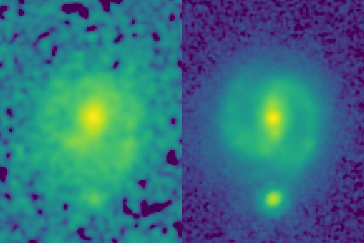
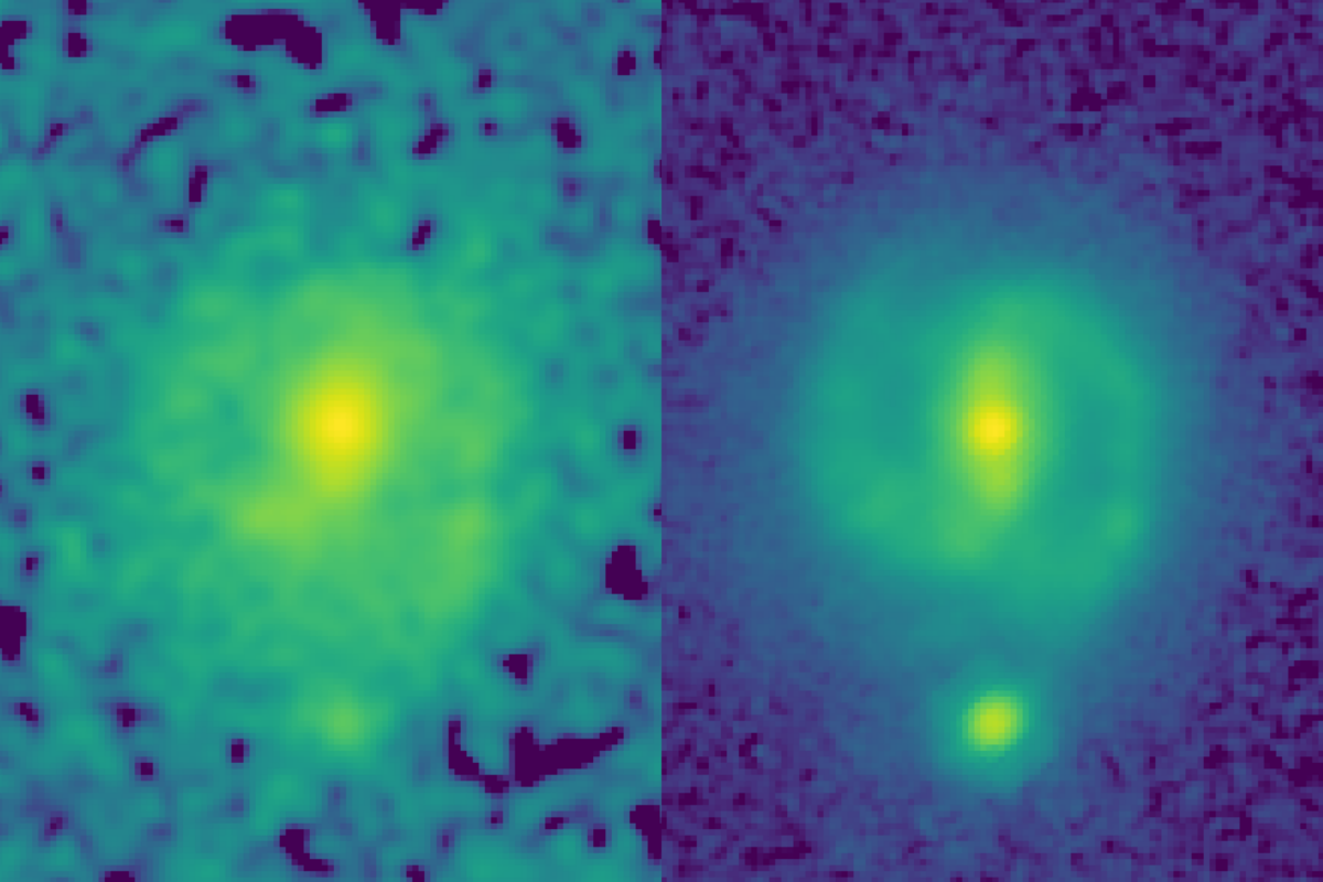
Prior to the James Webb Space Telescope (JWST), images from the Hubble Space Telescope had never detected bars at such young epochs. In a Hubble image, one galaxy, EGS-23205, is little more than a disk-shaped smudge, but in the corresponding JWST image taken this past summer, it’s a beautiful spiral galaxy with a clear stellar bar.

A side-by-side image of an image of a galaxy taken by the Hubble and James Webb telescopes.
The power of JWST to map galaxies at high resolution and at longer infrared wavelengths than Hubble allows it look through dust and unveil the underlying structure and mass of distant galaxies. This can be seen in these two images of the galaxy EGS23205, seen as it was about 11 billion years ago. In the HST image (left, taken in the near-infrared filter), the galaxy is little more than a disk-shaped smudge obscured by dust and impacted by the glare of young stars, but in the corresponding JWST mid-infrared image (taken this past summer), it’s a beautiful spiral galaxy with a clear stellar bar. (Credit: NASA/CEERS/UT Austin)

“I took one look at these data, and I said, ‘We are dropping everything else!’” says Shardha Jogee, professor of astronomy at the University of Texas at Austin.

“The bars hardly visible in Hubble data just popped out in the JWST image, showing the tremendous power of JWST to see the underlying structure in galaxies,” she says, describing data from the Cosmic Evolution Early Release Science Survey (CEERS), led by UT Austin professor, Steven Finkelstein.

“It’s like going into a forest that nobody has ever gone into.”

The team identified another barred galaxy, EGS-24268, also from about 11 billion years ago, which makes two barred galaxies existing farther back in time than any previously discovered.

In an article accepted for publication in The Astrophysical Journal Letters, they highlight these two galaxies and show examples of four other barred galaxies from more than 8 billion years ago.

“For this study, we are looking at a new regime where no one had used this kind of data or done this kind of quantitative analysis before,” says Yuchen “Kay” Guo, a graduate student who led the analysis, “so everything is new. It’s like going into a forest that nobody has ever gone into.”

Bars play an important role in galaxy evolution by funneling gas into the central regions, boosting star formation.

“Bars solve the supply chain problem in galaxies,” Jogee says. “Just like we need to bring raw material from the harbor to inland factories that make new products, a bar powerfully transports gas into the central region where the gas is rapidly converted into new stars at a rate typically 10 to 100 times faster than in the rest of the galaxy.”

Bars also help to grow supermassive black holes in the centers of galaxies by channeling the gas part of the way.

The discovery of bars during such early epochs shakes up galaxy evolution scenarios in several ways.

“This discovery of early bars means galaxy evolution models now have a new pathway via bars to accelerate the production of new stars at early epochs,” Jogee says.

And the very existence of these early bars challenges theoretical models as they need to get the galaxy physics right in order to predict the correct abundance of bars. The team will be testing different models in their next papers.

JWST can unveil structures in distant galaxies better than Hubble for two reasons: First, its larger mirror gives it more light-gathering ability, allowing it to see farther and with higher resolution. Second, it can see through dust better as it observes at longer infrared wavelengths than Hubble.

Undergraduate students Eden Wise and Zilei Chen played a key role in the research by visually reviewing hundreds of galaxies, searching for those that appeared to have bars, which helped narrow the list to a few dozen for the other researchers to analyze with a more intensive mathematical approach.

Additional coauthors are from UT Austin and other institutions in the US, the UK, Japan, Spain, France, Italy, Australia, and Israel.

Funding for this research came from, in part, the Roland K. Blumberg Endowment in Astronomy, the Heising-Simons Foundation, and NASA. This work relied on resources at the Texas Advanced Computing Center.

Source: UT Austin

source

Can hidden magnetic tags defeat counterfeit goods?

Researchers are using techniques from metal additive manufacturing to embed a hidden cache of information within products to help combat counterfeit goods.

Ensuring manufactured goods and components have not been copied and replaced illegally by counterfeit goods is a high-priority concern of the manufacturing and defense industries in the United States and around the world.

A potential solution could affect areas ranging from enhancing biomedical implants to protecting national defense assets.

Researchers from Texas A&M University have developed a method of imprinting a hidden magnetic tag, encoded with authentication information, within manufactured hardware during the part fabrication process. The process holds the potential to expose counterfeit goods more easily by replacing physical tags—such as barcodes or quick response (QR) codes—with these hidden magnetic tags, which serve as permanent and unique identifiers.

The team recently published its research in the journal Additive Manufacturing.

Ensuring security and reliable authentication in manufacturing is a critical national concern, with the US investing billions of dollars in manufacturing. Without such a method readily available, it can be nearly impossible to differentiate an authentic part or component from its counterfeit copy.

“The issue is that when I come up with an idea, device, or part, it is very easy for others to copy and even fabricate it much more cheaply—though maybe at a lower quality,” says Ibrahim Karaman, professor and department head of the materials science and engineering department. “Sometimes they even put the same brand name, so how do you make sure that item isn’t yours? [The embedded magnetic tag] gives us an opportunity and a new tool to make sure that we can protect our defense and manufacturing industries.”

The team is implementing metal additive manufacturing techniques to accomplish its goal of successfully embedding readable magnetic tags into metal parts without compromising performance or longevity. Researchers used 3D printing to embed these magnetic tags below the surface into nonmagnetic steel hardware.

Other applications for this method include traceability, quality control, and more, largely depending on the industry that uses it.

Once embedded into a nonmagnetic item, the magnetic tag is readable using a magnetic sensor device—such as a smartphone—by scanning near the correct location on the product.

While other methods exist for imprinting information, they primarily require sophisticated and costly equipment.

“Different approaches have been used to try to locally change the properties of the metals during the manufacturing process to be able to codify information within the part,” says Daniel Salas Mula, a researcher with the Texas A&M Engineering Experiment Station.

“This is the first time that magnetic properties of the material are being used in this way to introduce information within a nonmagnetic part, specifically for the 3D printing of metals.”

Doctoral student Deniz Ebeperi says that to map the magnetic reading of the part, the team created a custom three-axis magnetic sensor capable of mapping the surface and revealing the regions where the embedded magnetic tag was accessible.

While the system is more secure than a physical tag or code located on the exterior of an item, the team is still working to improve the complexity of the method’s security.

As the project continues, Karaman says the next steps include developing a more secure method of reading the information, possibly through the implementation of a physical “dual-authentication” requiring the user to apply a specific treatment or stimulus to unlock access to the magnetic tag.

Additional collaborators are from Texas A&M University and Purdue University. The project had support from the SecureAmerica Institute.

Source: Texas A&M University

source

Glassfrogs hide their blood to sleep in camouflage

Glassfrogs make themselves transparent while they rest by taking red blood cells from circulation and concealing them in their livers, research finds.

It’s easy to miss a glassfrog in its natural environment. The northern glassfrog, Hyalinobatrachium fleischmanni, measures no more than a few centimeters, and they are most active at night, when their green skin helps them blend in with the surrounding leaves and foliage.

But these amphibians become true masters of camouflage during the day when they’re asleep.

shapes of two frogs barely visible through leaf
Sleeping mated glassfrogs. (Credit: Duke)

“When glassfrogs are resting, their muscles and skin become transparent, and their bones, eyes, and internal organs are all that’s visible,” says Carlos Taboada, a postdoctoral fellow at Duke University and a co-first author of the paper. “These frogs sleep on the bottoms of large leaves, and when they’re transparent, they can perfectly match the colors of the vegetation.”

Many animals in the sea can change the color of their skin or become completely transparent, but it’s a far less common skillset on land. One reason transparency is so difficult to achieve is because of red blood cells in the circulatory system. Red blood cells are adept at absorbing green light, which is the color of light usually reflected by plants and other vegetation. In return, these oxygen-rich cells reflect red light, making blood—and by extension the circulatory system—highly visible, especially against a bright green leaf.

Glassfrogs are some of the only land-based vertebrates that can achieve transparency, which has made them a target for study. Taboada first began studying glass frogs as a postdoctoral fellow in the lab of Sönke Johnsen, a professor of biology at Duke who specializes in studying transparency. Working with Jesse Delia, who traveled around the world collecting different glassfrogs for the study, they observed that red blood cells seemed to be disappearing from the circulating blood whenever the frogs became transparent.

They conducted additional imaging tests on the animals, proving via optical models that the animals were able to achieve transparency because they were pushing red blood cells out of their vessels. He suspected that the cells were being stored in one of the frog’s inner organs which are packaged in a reflective membrane. The finding appears in the journal Science.

For a see-through animal, its biology was shockingly challenging to decipher. The research drew on the expertise of biologists and biomedical engineers at Duke and at the American Museum of Natural History, Stanford University, and the University of Southern California.

“If these frogs are awake, stressed, or under anesthesia their circulatory system is full of red blood cells and they are opaque,” explains Delia, now a postdoctoral fellow at the American Museum of Natural History. “The only way to study transparency is if these animals are happily asleep, which is difficult to achieve in a research lab. We were really banging our heads against the wall for a solution.”

But Taboada had learned about an imaging technology called photoacoustic microscopy, or PAM, when he was studying biliverdin, the compound that gives certain species of frogs their signature green color. PAM involves shooting a safe laser beam of light into tissue, which is then absorbed by molecules and converted into ultrasonic waves. These sound waves are then used to make detailed biomedical images of the molecules. The imaging tool is non-invasive, quiet, sensitive, and, in a stroke of luck, available at Duke.

“PAM is the ideal tool for non-invasive imaging of red blood cells because you don’t need to inject contrast agents, which would be very difficult for these frogs,” explains Junjie Yao, an assistant professor of biomedical engineering at Duke who specializes in PAM technologies. “The red blood cells themselves provide the contrast, because different types of cells absorb and reflect different wavelengths of light. We could optimize our imaging systems to specifically look for red blood cells and track how much oxygen was circulating in the frog’s bodies.”

In their imaging set-up, the frogs slept upside down in a petri dish, similar to how they would sleep on a leaf, and the team shined a green laser at the animal. The red blood cells in the frog’s body absorbed the green light and emitted ultrasonic waves, which were then picked up by an acoustic sensor to trace their whereabouts, with high spatial resolution and high sensitivity.

The results were startlingly clear: When the frogs were asleep, they removed nearly 90% of their circulating red blood cells and stored them in their liver.

In further tests, the team also saw that red blood cells flowed out of the liver and circulated when the frogs were active, and then re-aggregated in the liver while the frogs were recovering.

“The primary result is that whenever glassfrogs want to be transparent, which is typically when they’re at rest and vulnerable to predation, they filter nearly all the red blood cells out of their blood and hide them in a mirror-coated liver—somehow avoiding creating a huge blood clot in the process,” says Johnsen. “Whenever the frogs need to become active again, they bring the cells back into the blood stream, which gives them the metabolic capacity to move around.”

According to Delia and Taboada, this process raises questions about how the frogs can safely store almost all their red blood cells in their liver without clotting or damaging their peripheral tissues.

One potential next step, they say, could be to study this mechanism and how it could one day apply to vascular issues in humans.

This work also introduces glassfrogs as a useful model for research, especially when paired with the state-of-the-art photoacoustic imaging. As long-time glassfrog researchers, they are excited about the new avenues of study now available to them and interested collaborators.

“We can learn more about the glassfrog’s physiology and behavior, or we can use these models to optimize imaging tools for biomedical engineering,” Delia says.

This work had support from National Geographic Society; the Human Frontier Science Program postdoctoral fellowship; the Gerstner Scholars Fellowship provided by the Gerstner Family Foundation and the Richard Gilder Graduate School at the American Museum of Natural History; start-up funds from Stanford University and start-up funds from Duke University; the National Institutes of Health; the National Science Foundation CAREER Award; the Duke Institute of Brain Science Incubator award; the American Heart Association Collaborative Sciences award; and the Chan Zuckerberg Initiative.

Source: Duke University

source

Expert: TikTok could be a risk to national security

Although TikTok users consider the app harmless fun, a growing number of cybersecurity experts and elected officials aren’t so sure.

More than 86 million Americans use the social media app TikTok to create, share, and view short videos, featuring everything from cute animals and influencer advice to comedy and dance performances.

Concerned experts point out that TikTok’s parent company, the Beijing-based ByteDance, has been accused of working with the Chinese government to censor content and could also collect sensitive data on users.

To date, at least 14 of the United States have enacted legislation or created rules blocking government computers’ access to the app, and a bipartisan bill introduced in December in Congress seeks a ban on the app for all US users.

Christopher Wray, FBI director, spoke publicly on the issue last month, warning that control of the popular social media app is in the “hands of a government that doesn’t share our values.”

Cybersecurity expert Anton Dahbura, executive director of the Johns Hopkins University Information Security Institute, discusses the issue here:

source

Tweets could track the spread of invasive insects

Twitter and online news articles show potential for tracking the timing and location of invasive insect spread, a new study shows.

The researchers say these sources are promising for filling in gaps when official data are not widely available.

“The idea was to explore if we could use this data to fill in some of the information gaps about pest spread, and ultimately, to support the development of better predictive models of where pest spread is happening, and when to use costly control measures,” says Laura Tateosian, associate teaching professor in the Center for Geospatial Analytics at North Carolina State University.

“Even though these are not formal scientific sources, we found that we could clearly see some of the major events that were occurring about two invasive pests in the news, and on Twitter.”

In the study, the researchers tracked past tweets about two insects—the spotted lanternfly and Tuta absoluta—compiled by a internet-based subscription service, Brandwatch, as well as online news articles aggregated by Google News and GDELT, or the Global Database of Events, Language, and Tone Project.

The spotted lanternfly, which was first reported in the United States in Pennsylvania in 2014, is an insect native to Asia that can damage or destroy grapes, cherries, hops, certain lumber trees, and other plants. The research team tracked historical posts about spotted lanternfly in Pennsylvania in a single year in 2017, and then globally between 2011 and 2021.

Tuta absoluta, an insect also known as the tomato leaf miner, is native to South America. It was discovered in Spain in 2006, and has spread into parts of Europe, Africa, Asia, and the Middle East. It has been nicknamed the “tomato Ebola” because of the devastation it can cause to tomato crops. The researchers tracked posts about Tuta absoluta between 2011 and 2021.

“While some invasive insects have reached their global range, in both of these cases, the pests are actively spreading,” says Ariel Saffer, a graduate student in geospatial analytics.

“We launched this as a proof-of-concept study to see if it would be scientifically reasonable to use these sources to track pest spread. We compared information in places where the insects were known to be present to see if these sources accurately captured existing knowledge.”

The researchers found that activity on Twitter and in news stories tracked some of the patterns in official surveys. For example, the volume of Twitter posts and news activity about spotted lanternfly tracked the seasonal pest cycle, with more activity in the summer and fall.

In terms of location, they saw a high volume of tweets and news articles in areas located at the epicenter of outbreaks. In Pennsylvania, news articles captured a subset of counties confirmed in 2017 by USDA Animal and Plant Health Inspection Service survey data, but also uncovered one county not listed in official records.

For Tuta absoluta, the team found posts on Twitter and in news stories often coincided with global pest spread, as compared to reports gathered by the European and Mediterranean Plant Protection Organization (EPPO). Information in news and Twitter posts also aligned with survey data for this pest in Nigeria, and sometimes before that information was widely available in scientific sources.

The findings suggest Twitter and news information could be useful to supplement official data sources, but more work is needed, the researchers say.

“News media and social media have the potential to give you more immediate insight into what’s going on, especially if scientific information on insect spread is not immediately published in scientific literature, or not widely available to other scientists,” Saffer says.

“Also, relying on data from scientific publications can sometimes offer a patchwork coverage of space and time, depending on when that study happened. It can be hard to get aggregated information in continuous time, especially at the global scale, as that information can be managed by multiple agencies.”

The study appears in the journal Computers, Environment and Urban Systems.

Source: NC State

source

Common fatty acid fuels psoriasis pain, but not itch

A common fatty acid found in the Western diet breaks down into compounds that contribute to increased temperature and pain—but not itch—sensitivity in people with psoriasis lesions, researchers report.

The finding could lead to better understanding of how lipids communicate with sensory neurons, and potentially to improved pain and sensitivity treatments for psoriasis patients.

Linoleic acid is a fatty acid found in vegetable oils, nuts, and seeds, and is one of the predominant fatty acids found in the Western diet. Metabolites from linoleic acid—the products formed when the body breaks it down through digestion—play a role in skin barrier function.

“We noticed high levels of two types of lipids derived from linoleic acid in psoriatic lesions,” says Santosh Mishra, associate professor of neuroscience at North Carolina State University and corresponding author of the research in JID Innovations.

“That led us to wonder whether the lipids might affect how sensory neurons in these lesions communicate. We decided to investigate whether their presence could be related to the temperature or pain hypersensitivity that many psoriasis patients report.”

The research team used mass spectrometry to create lipid profiles of skin from psoriatic lesions. They focused on two types of linoleic acid-derived lipids, or oxylipids: 13-hydroxy-9,10-epoxy octadecenoate (9,13-EHL) and 9,10,13-trihydroxy-octadecenoate (9,10,13-THL). The first form, 9,13-EHL, can convert into the more stable 9,10,13-THL form via interaction with certain enzymes.

The researchers found that while both forms bind to receptors on sensory neurons within the skin, the more stable form—9,10,13-THL—had a longer lasting effect than 9,13-EHL.

They also found that once the lipids bind to the neuronal receptor, they activate the neurons expressing TRPA1 and TRPV1 receptors that are involved in temperature and pain hypersensitivity, opening communications channels to the central nervous system.

Interestingly, the lipids did not have any effect on itch.

“It was surprising that these lipids could create hypersensitivity but not impact itch sensation, which is usually the most troublesome symptom associated with psoriasis,” Mishra says. “This most likely has to do with how the neuron is activated—a mechanism we still haven’t uncovered.”

Now that an association between linoleic acid and hypersensitivity to temperature and pain has been established, the researchers want to further explore exactly how this response is created. They hope that the answers may lead to solutions that can relieve these symptoms in psoriasis patients.

“We know that this lipid moves from one form to another, but don’t yet know what causes that,” Mishra says. “We also know what protein the lipids are binding to, but not where the bond occurs. Answering these questions may hopefully lead to new therapies—or dietary solutions— for some psoriasis sufferers.”

The National Institute on Aging and the National Institutes of Health supported the work.

Source: NC State

source

Simple nasal swab finds sneaky viruses other tests miss

Testing for the presence of a single immune system molecule on nasal swabs can help detect stealthy viruses not identified in standard tests, a new study shows.

As the COVID-19 pandemic showed, potentially dangerous new viruses can begin to spread in the population well before the global public health surveillance system can detect them.

“Finding a dangerous new virus is like searching for a needle in a haystack,” says Ellen Foxman, associate professor of laboratory medicine and immunobiology at Yale University and senior author of the study in The Lancet Microbe. “We found a way to significantly reduce the size of the haystack.”

Public health officials typically look to a few sources for warning signs of emerging disease. They study emerging viruses in animals that may transmit the infection to humans.

But determining which of the hundreds, or thousands, of new viral variants represent a true danger is difficult. And they look for outbreaks of unexplained respiratory ailments, which was how SARS-Cov-2, the virus that causes COVID-19, was discovered in China late in 2019.

By the time an outbreak of a novel virus occurs, however, it may be too late to contain its spread.

For the new study, Foxman and her team revisited an observation made in her lab in 2017, which they thought may provide a new way to monitor for unexpected pathogens.

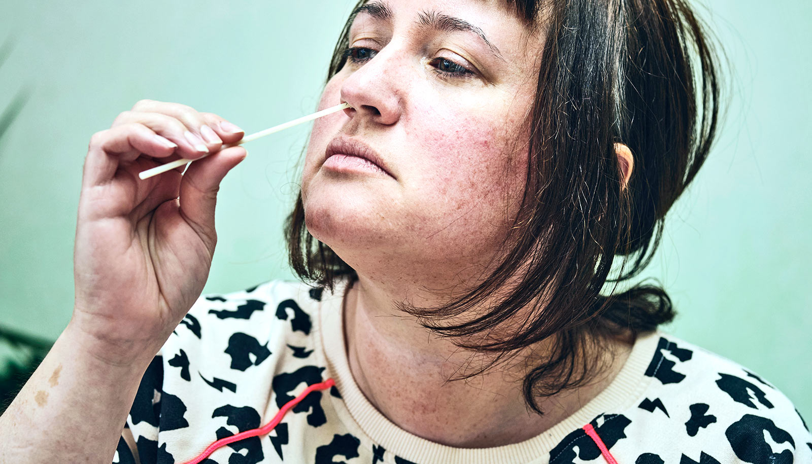
Nasal swabs are commonly taken from patients with suspected respiratory infections and are tested to detect specific signatures of 10 to 15 known viruses. Most tests come back negative.

But as Foxman’s team observed in 2017, in a few cases the swabs of those who tested negative for the “usual suspect” viruses still exhibited signs that antiviral defenses were activated, indicating the presence of a virus. The telltale sign was a high level of a single antiviral protein made by the cells that line the nasal passages.

Based on that finding, the researchers applied comprehensive genetic sequencing methods to old samples containing the protein and, in one sample, found an unexpected influenza virus, called influenza C.

The researchers also used this same strategy of retesting old samples to search for missed cases of COVID-19 during the first two weeks of March 2020. While cases of the virus had surfaced in New York State around that same time, testing was not readily available until weeks later.

Hundreds of nasal swab samples collected from patients at Yale-New Haven Hospital during that time had tested negative for standard signature viruses. When tested for the immune system biomarker, the vast majority of those samples showed no trace of activity of the antiviral defense system. But a few did; among those, the team found four cases of COVID-19 that had gone undiagnosed at the time.

The findings reveal that testing for an antiviral protein made by the body, even if the tests for known respiratory viruses are negative, can help pinpoint which nasal swabs are more likely to contain unexpected viruses.

Specifically, screening for the biomarker can allow researchers to narrow down the search for unexpected pathogens, making it feasible to do surveillance for unexpected viruses using swabs collected during routine patient care.

Samples found to possess the biomarker can be analyzed using more complex genetic testing methods to identify unexpected or emerging pathogens circulating in the patient population and jumpstart a response from the health care community.

Yale’s Nagarjuna R. Cheemarla and Jason Bishai are co-lead authors of the paper, as are former Yale researchers Amelia Hanron and Joseph R. Fauver.

Source: Yale University

source

Stretchy sensor works for health and video games

Researchers have developed a stretchable strain sensor that has an unprecedented combination of sensitivity and range.

That allows it to detect even minor changes in strain with greater range of motion than previous technologies.

The researchers demonstrated the sensor’s utility by creating new health monitoring and human-machine interface devices.

Strain is a measurement of how much a material deforms from its original length. For example, if you stretched a rubber band to twice its original length, its strain would be 100%.

“And measuring strain is useful in many applications, such as devices that measure blood pressure and technologies that track physical movement,” says Yong Zhu, corresponding author of a paper on the work and professor of mechanical and aerospace engineering at North Carolina State University.

“But to date there’s been a trade-off. Strain sensors that are sensitive—capable of detecting small deformations—cannot be stretched very far. On the other hand, sensors that can be stretched to greater lengths are typically not very sensitive. The new sensor we’ve developed is both sensitive and capable of withstanding significant deformation,” says Zhu.

“An additional feature is that the sensor is highly robust even when over-strained, meaning it is unlikely to break when the applied strain accidentally exceeds the sensing range.”

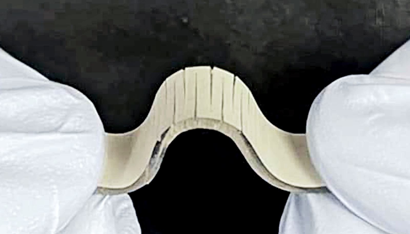
The new sensor consists of a silver nanowire network embedded in an elastic polymer. The polymer features a pattern of parallel cuts of a uniform depth, alternating from either side of the material: one cut from the left, followed by one from the right, followed by one from the left, and so on.

“This feature—the patterned cuts—is what enables a greater range of deformation without sacrificing sensitivity,” says Shuang Wu, who is first author of the paper and a recent PhD graduate.

The sensor measures strain by measuring changes in electrical resistance. As the material stretches, resistance increases. The cuts in the surface of the sensor are perpendicular to the direction that it is stretched. This does two things. First, the cuts allow the sensor to deform significantly. Because the cuts in the surface pull open, creating a zigzag pattern, the material can withstand substantial deformation without reaching the breaking point. Second, when the cuts pull open, this forces the electrical signal to travel further, traveling up and down the zigzag.

“To demonstrate the sensitivity of the new sensors, we used them to create new wearable blood pressure devices,” Zhu says. “And to demonstrate how far the sensors can be deformed, we created a wearable device for monitoring motion in a person’s back, which has utility for physical therapy.”

“We have also demonstrated a human-machine interface,” Wu says. “Specifically, we used the sensor to create a three-dimensional touch controller that can be used to control a video game.”

“The sensor can be easily incorporated into existing wearable materials such as fabrics and athletic tapes, convenient for practical applications,” Zhu says. “And all of this is just scratching the surface. We think there will be a range of additional applications as we continue working with this technology.”

The paper appears in the journal ACS Applied Materials & Interfaces.

Support for the work came from the National Science Foundation, the National Institutes of Health, and the US Department of Defense.

Source: NC State

source

Dry eye changes how injured cornea heals itself

A new study with mice finds that proteins made by stem cells that regenerate the cornea may be new targets for treating and preventing injuries.

People with a condition known as dry eye disease are more likely than those with healthy eyes to suffer injuries to their corneas.

Dry eye disease occurs when the eye can’t provide adequate lubrication with natural tears. People with the common disorder use various types of drops to replace missing natural tears and keep the eyes lubricated, but when eyes are dry, the cornea is more susceptible to injury.

“We have drugs, but they only work well in about 10% to 15% of patients,” says senior investigator Rajendra S. Apte, professor in the department of ophthalmology and visual sciences at Washington University in St. Louis.

“In this study involving genes that are key to eye health, we identified potential targets for treatment that appear different in dry eyes than in healthy eyes.

“Tens of millions of people around the world—with an estimated 15 million in the United States alone—endure eye pain and blurred vision as a result of complications and injury associated with dry eye disease, and by targeting these proteins, we may be able to more successfully treat or even prevent those injuries.”

For the study in the Proceedings of the National Academy of Sciences, the researchers analyzed genes expressed by the cornea in several mouse models—not only of dry eye disease, but also of diabetes and other conditions. They found that in mice with dry eye disease, the cornea activated expression of the gene SPARC. They also found that higher levels of SPARC protein were associated with better healing.

“We conducted single-cell RNA sequencing to identify genes important to maintaining the health of the cornea, and we believe that a few of them, particularly SPARC, may provide potential therapeutic targets for treating dry eye disease and corneal injury,” says first author Joseph B. Lin, an MD/PhD student in Apte’s lab.

“These stem cells are important and resilient and a key reason corneal transplantation works so well,” Apte explains. “If the proteins we’ve identified don’t pan out as therapies to activate these cells in people with dry eye syndrome, we may even be able to transplant engineered limbal stem cells to prevent corneal injury in patients with dry eyes.”

The National Eye Institute, the National Institute of Diabetes and Digestive and Kidney Diseases, and the National Institute of General Medical Sciences of the National Institutes of Health supported the work. Additional funding came from the Jeffrey T. Fort Innovation Fund, a Centene Corp. contract for the Washington University-Centene ARCH Personalized Medicine Initiative, and Research to Prevent Blindness.

Source: Washington University in St. Louis

source

Study may explain why too much of a good smell can stink

New research reveals an added layer of nuance in our sense of smell.

The delicate fragrance of jasmine is a delight to the senses. The sweet scent is popular in teas, perfumes and potpourri. But take a whiff of the concentrated essential oil, and the pleasant aroma becomes almost cloying.

Part of the flower’s smell actually comes from the compound skatole, a prominent component of fecal odor.

“Consider for instance the smell of a ripe banana from a distance (sweet and fruity) versus up-close (overpowering and artificial).”

Our sense of smell is clearly a complex process; it involves hundreds of different odorant receptors working in concert. The more an odor stimulates a particular neuron, the more electrical signals that neuron sends to the brain.

But the new research reveals that these neurons actually fall silent when an odor rises above a certain threshold. Remarkably, this was integral to how the brain recognized each smell.

“It’s a feature; it’s not a bug,” says Matthieu Louis, an associate professor in the department of molecular, cellular, and developmental biology at the University of California, Santa Barbara.

The paradoxical finding, published in Science Advances, shakes up our understanding of olfaction.

“The same odor can be represented by very different patterns of active olfactory sensory neurons at different concentrations,” Louis says. “This might explain why some odors can be perceived as very different to us at low, medium, and very high concentrations. Consider for instance the smell of a ripe banana from a distance (sweet and fruity) versus up-close (overpowering and artificial).”

Humans have several million sensory neurons in our noses, and each of these has one type of odorant receptor. Altogether, we have about 400 different types of receptors with overlapping sensitivity. Each chemical compound is like a different shoe that the receptor is trying on. Some shoes fit snugly, some fit well, while others don’t fit at all. A better fit produces a stronger response from the receptor. Increasing an odor’s concentration recruits neurons with receptors that have are less sensitive to that substance. Our brain uses the combination of activated neurons to distinguish between odors.

Scientists thought that neurons would effectively max out above certain odor concentrations, at which point their activity would plateau. But the team led by Louis’ graduate student, David Tadres, found the exact opposite: Neurons actually fall silent above a certain level, with the most sensitive ones dropping off first.

Looking at flies

Fruit fly larvae make an ideal model for studying olfaction. They have as many types of odorant receptors as the number of sensory neurons—namely, 21. This one-to-one correspondence makes it simple to test what each neuron is doing.

For the study, Tadres examined larvae with a mutation that entirely eliminated their sense of smell. He then selectively turned that sense back on in a single sensory neuron, enabling the larvae to detect only odors that activated that specific receptor. He placed them next to an odor source and watched.

Even with a single functioning olfactory channel, the larvae could still move toward the stronger smell. But remarkably, they stopped a certain distance away from the source, and just circled it in a fixed orbit. Tadres repeated the experiment with a neuron slightly less sensitive to the odor he was testing, and found that the larvae got closer to the source before stopping.

Puzzled by this behavior, Tadres used electrodes to measure the activity of the sensory neuron. As expected, signaling increased as the odor became more concentrated. But rather than plateau above a certain level, the activity crashed to zero. That’s why the mutant larvae circled the odor source; above a certain concentration, the smell simply disappeared.

“The silencing of the olfactory sensory neuron could easily explain the circling behavior, which was mysterious before,” Tadres says. “From there it wasn’t hard to extrapolate that the current view of how odors are encoded at different concentrations needed to be updated.”

Researchers knew that excessive stimulation can cause nerves to go silent, an effect called “depolarization block.” However, the consensus was that this sort of overload doesn’t occur under natural, healthy conditions. Indeed, this response is associated with issues like epilepsy when it occurs in the central brain. But when Tadres observed it affecting the larvae’s behavior, he suspected that it wasn’t merely an artifact of the experiment.

Digging deeper

Tadres and Louis began investigating the cause of the depolarization block. For assistance, they reached out to Professor Jeff Moehlis, chair of the mechanical engineering department, and Louis’ doctoral student Philip Wong (co-advised by Moehlis), who started constructing a mathematical model of the system.

The voltage across a neuron’s membrane can be described by a system of equations. This model was a breakthrough finding in 1952, and earned a Nobel Prize for its discoverers, Alan Hodgkin and Andrew Huxley. For this case study, Wong added a mathematical representation of the odorant receptor, the “trigger” that initiates the rest of the model. He also included a modification from the field of epilepsy research wherein high stimulation turns off certain ion channels in the cell membrane, preventing a neuron from firing.

Wong’s model was able to fit and predict Tadres’ measurements of the neuron’s electrical activity. “This was quite useful because the electrophysiology data was difficult to collect and very time consuming to analyze,” Wong says.

In addition to corroborating the experimental results, the model is guiding the team as they continue investigating this effect. “This model may tell us exactly how each neuron is responding to different odors,” Wong says.

The model’s success points to a possible source of the depolarization block: a specific ion channel present in neurons across the animal kingdom. If true, this suggests that most sensory neurons might fall silent following strong and sustained stimulation. The team hopes to validate this hypothesis in an upcoming study.

What’s more, the model predicted that the system would behave differently going up from low odor concentrations versus coming down from high concentrations. Measuring the voltage of the larvae’s neurons confirmed this. When going down, the neuron did not reactivate below the threshold where it had fallen silent. In fact, it largely remained silent until the odor concentration came back down to zero before returning to normal activity.

Our complex sense of smell

This study demonstrated that high odor concentrations can silence the most sensitive receptors. This counterintuitive result marks a fundamental shift in our understanding of smell.

“As you increase the concentration of an odor, you’ll start recruiting more and more odorant receptors that aren’t as sensitive to that compound,” Louis explains. “And so, the common view until our work was that you just kept adding active odorant receptors to the picture.”

This makes sense, until you consider the system as a whole. If this were the case, then a compound should activate pretty much all of the receptors above a certain level. “So it would be impossible for you to distinguish between two different odors at very high concentrations,” Tadres says. “And that’s clearly not the case.”

Having certain sensory neurons drop out as others join in might help preserve the distinction between odors at high concentrations. And this could prove important for survival. It might prevent poisons and nutrients that share certain compounds from smelling alike when you take a big whiff of them.

It could also have consequences for how we perceive odors. “We speculate that removing successive high-sensitivity olfactory sensory neurons is like removing the root of a musical chord,” Louis says. “This omission of the root is going to alter the way your brain perceives the chord associated with a set of notes. It’s going to give it a different meaning.”

A subtle floral note suggests an orchard may be in bloom nearby, useful information for a hungry animal. Meanwhile, the same compounds in higher concentrations could produce the pungent ripeness of decaying fruit or even sewage: something best avoided. Studies like this reveal ever more complexities to our sense of smell, which evolved to help us navigate an equally complex chemical landscape.

Source: UC Santa Barbara
source